Journal of Molecular and Cellular Cardiology ( IF 4.9 ) Pub Date : 2020-06-06 , DOI: 10.1016/j.yjmcc.2020.05.021 Zhen-Ye Zhang 1 , Ling-Ling Qian 1 , Ning Wang 1 , Ling-Feng Miao 1 , Xin Ma 2 , Shi-Peng Dang 1 , Ying Wu 1 , Xiao-Yu Liu 1 , Xiao-Yan Li 1 , Qiang Chai 3 , Min Pan 4 , Fu Yi 5 , Tian-You Ling 6 , Ru-Xing Wang 1
|
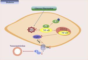
Glucose fluctuations may contribute to large conductance calcium activated potassium (BK) channel dysfunction. However, the underlying mechanisms remain elusive. The aim of this study was to investigate the molecular mechanisms involved in BK channel dysfunction as a result of glucose fluctuations. A rat diabetic model was established through the injection of streptozotocin. Glucose fluctuations in diabetic rats were induced via consumption and starvation. Rat coronary arteries were isolated and coronary vascular tensions were measured after three weeks. Rat coronary artery smooth muscle cells were isolated and whole-cell BK channel currents were recorded using a patch clamp technique. Human coronary artery smooth muscle cells in vitro were used to explore the underlying mechanisms. After incubation with iberiotoxin (IBTX), the Δ tensions (% Max) of rat coronary arteries in the controlled diabetes mellitus (C-DM), the uncontrolled DM (U-DM) and the DM with glucose fluctuation (GF-DM) groups were found to be 84.46 ± 5.75, 61.89 ± 10.20 and 14.77 ± 5.90, respectively (P < .05), while the current densities of the BK channels in the three groups were 43.09 ± 4.35 pA/pF, 34.23 ± 6.07 pA/pF and 17.87 ± 4.33 pA/pF, respectively (P < .05). The Δ tensions (% Max) of rat coronary arteries after applying IBTX in the GF-DM rats injected with 0.9% sodium chloride (NaCl) (GF-DM + NaCl) and the GF-DM rats injected with N-acetyl-L-cysteine (NAC) (GF-DM + NAC) groups were found to be 8.86 ± 1.09 and 48.90 ± 10.85, respectively (P < .05). Excessive oxidative stress and the activation of protein kinase C (PKC) α and nuclear factor (NF)-κB induced by glucose fluctuations promoted the decrease of BK-β1 expression, while the inhibition of reactive oxygen species (ROS), PKCα, NF-κB and muscle ring finger protein 1 (MuRF1) reversed this effect. Glucose fluctuations aggravate BK channel dysfunction via the ROS overproduction and the PKCα/NF-κB/MuRF1 signaling pathway.
中文翻译:
葡萄糖波动通过 PKCα/NF-κB/MuRF1 信号传导促进血管 BK 通道功能障碍。
葡萄糖波动可能导致大电导钙激活钾 (BK) 通道功能障碍。然而,潜在的机制仍然难以捉摸。本研究的目的是研究由于葡萄糖波动导致 BK 通道功能障碍的分子机制。通过注射链脲佐菌素建立大鼠糖尿病模型。糖尿病大鼠的葡萄糖波动是通过消耗和饥饿引起的。三周后分离大鼠冠状动脉并测量冠状血管张力。分离大鼠冠状动脉平滑肌细胞并使用膜片钳技术记录全细胞 BK 通道电流。体外人冠状动脉平滑肌细胞被用来探索其潜在机制。与伊比利亚毒素 (IBTX) 孵育后,控制糖尿病 (C-DM)、未控制糖尿病 (U-DM) 和血糖波动糖尿病 (GF-DM) 组大鼠冠状动脉的 Δ 张力 (% Max)分别为 84.46 ± 5.75、61.89 ± 10.20 和 14.77 ± 5.90 ( P < .05),而三组中 BK 通道的电流密度为 43.09 ± 4.35 pA/pF,34.2073 pA/pF和 17.87 ± 4.33 pA/pF,分别为 ( P < .05)。GF-DM 大鼠注射 0.9% 氯化钠 (NaCl) (GF-DM + NaCl) 和注射 N-乙酰-L-的 GF-DM 大鼠应用 IBTX 后大鼠冠状动脉的 Δ 张力 (% Max)半胱氨酸 (NAC) (GF-DM + NAC) 组分别为 8.86 ± 1.09 和 48.90 ± 10.85 ( P < .05)。葡萄糖波动引起的过度氧化应激和蛋白激酶C(PKC)α和核因子(NF)-κB的激活促进了BK-β1表达的降低,而活性氧(ROS)、PKCα、NF- κB 和肌肉无名指蛋白 1 (MuRF1) 逆转了这种效应。葡萄糖波动通过 ROS 过量产生和 PKCα/NF-κB/MuRF1 信号通路加重 BK 通道功能障碍。


 京公网安备 11010802027423号