Annals of Anatomy ( IF 2.0 ) Pub Date : 2020-06-05 , DOI: 10.1016/j.aanat.2020.151549 Lorenzo Alibardi 1
|
Background
The regenerating blastema of the tail in the lizard Podarcis muralis contains numerous macrophages among the prevalent mesenchymal cells. Some macrophages are phagocytic but others are devoid of phagosomes suggesting that they have other roles aside phagocytosis.
Methods
The presence of healing macrophages (M2-like) has been tested using autoradiographic, immunohistochemical and ultrastructural studies.
Results
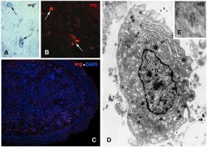
Autoradiography shows an uptake of tritiated arginine in sparse cells of the blastema and in the regenerating epidermis. Bioinformatics analysis suggests that epitopes for arginase-1 and -2, recognized by the employed antibody, are present in lizards. Immunofluorescence shows sparse arginase immunopositive macrophages in the blastema and few macrophages also in the apical wound epidermis. The ultrastructural study shows that macrophages contain dense secretory granules, most likely inactive lysosomes, and small cytoplasmic pale vesicles. Some of the small vesicles are arginase-positive while immunolabeling is very diffuse in the macrophage cytoplasm.
Conclusions
The presence of cells incorporating arginine and of arginase 1-positive cells suggests that M2-like macrophages are present among mesenchymal and epidermal cells of the regenerative tail blastema. M2-like macrophages may promote tail regeneration differently from the numerous pro-inflammatory macrophages previously detected in the scarring limb. The presence of M2-like macrophages in addition to hyaluronate, support the hypothesis that the regenerative blastema of the tail in lizards is an immuno-privileged organ where cell proliferation and growth occur without degenerating in a tumorigenic outgrowth.
中文翻译:
放射自显影和免疫标记表明,蜥蜴胚细胞含有精氨酸酶阳性的M2样巨噬细胞,可能支持尾巴再生。
背景
蜥蜴Podarcis muralis尾巴的再生胚细胞在普遍的间充质细胞中包含大量巨噬细胞。一些巨噬细胞具有吞噬功能,但其他巨噬细胞不含吞噬体,这表明它们除了吞噬作用外还具有其他作用。
方法
已使用放射自显影,免疫组织化学和超微结构研究测试了治愈性巨噬细胞(类M2)的存在。
结果
放射自显影显示在胚泡的稀疏细胞和再生表皮中摄取ti化的精氨酸。生物信息学分析表明,蜥蜴中存在被所用抗体识别的精氨酸酶-1和-2的表位。免疫荧光显示胚泡中的稀疏精氨酸酶免疫阳性巨噬细胞,并且在顶端伤口表皮中也只有少数巨噬细胞。超微结构研究表明,巨噬细胞含有致密的分泌颗粒,极有可能是无活性的溶酶体,以及小的胞质苍白囊泡。一些小囊泡精氨酸酶阳性,而免疫标记在巨噬细胞胞质中非常分散。
结论
掺入精氨酸的细胞和精氨酸酶1阳性细胞的存在表明M2样巨噬细胞存在于再生尾巴胚细胞的间充质和表皮细胞之间。M2类巨噬细胞可能促进尾巴再生,这不同于先前在疤痕肢中发现的众多促炎性巨噬细胞。除了透明质酸盐外,M2样巨噬细胞的存在还支持了这样的假说:蜥蜴尾巴的再生胚细胞是一种免疫特权的器官,在该器官中,细胞增殖和生长发生而在致瘤性生长中没有退化。


 京公网安备 11010802027423号