当前位置:
X-MOL 学术
›
Microsc. Res. Tech.
›
论文详情
Our official English website, www.x-mol.net, welcomes your
feedback! (Note: you will need to create a separate account there.)
Anatomical and histological studies of the alimentary canal of adult maize leaf weevil, Tanymecusdilaticollis Gyllenhal, 1834 (Coleoptera: Curculionidae).
Microscopy Research and Technique ( IF 2.0 ) Pub Date : 2020-06-01 , DOI: 10.1002/jemt.23507 Selami Candan 1 , Nurcan Özyurt Koçakoğlu 1 , Mustafa Güllü 2 , Üzeyir Çağlar 3
Microscopy Research and Technique ( IF 2.0 ) Pub Date : 2020-06-01 , DOI: 10.1002/jemt.23507 Selami Candan 1 , Nurcan Özyurt Koçakoğlu 1 , Mustafa Güllü 2 , Üzeyir Çağlar 3
Affiliation
|
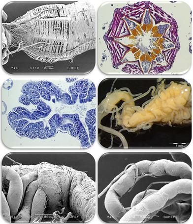
In this study, ananatomical and histological study was conducted on the alimentary canal of Tanymecusdilaticollis (Coleoptera: Curculionidae), which is an economic polyphagous pest species, to study the relationship between the structure of the alimentary canal and the feeding habit. Therefore, the structure of the alimentary canal of T. dilaticollis was examined using light and electron microscopies. Results have shown that the alimentary canal in T. dilaticollis is consisted of three separate regions as foregut, midgut, and hindgut structurally between the mouth and the anus, which pass from head, thorax, and abdomen. The foregut consists of pharynx, esophagus, crop and proventriculus and in the crop part, expansion is seen compared to other foregut parts. Midgut of T. dilaticollis is the largest part of digestion system. The anterior region of midgut is twofolds wider than the posterior region. The posterior midgut extends tubularly and it is connected to eightgastric caeca. The hindgut of T. dilaticollis consists of fourparts as pylorus, ileum, colon, and rectum. Well‐developed muscle layers are found near the rectum and genital chamber. These results contribute to further studies on the ecology and biological control agents of Coleoptera and to provide a broad comparison of alimentary canal of Coleoptera species.
中文翻译:

成年玉米叶片象鼻虫消化道的解剖学和组织学研究,Tanymecusdilaticollis Gyllenhal,1834年(鞘翅目:Curculionidae)。
在这项研究中,对一种经济多食性害虫种-食虫科(Tanymecusdilaticollis,Coleoptera:Curculionidae)的消化道进行了解剖学和组织学研究,以研究消化道的结构与喂养习惯之间的关系。因此,使用光和电子显微镜检查了T. diticicollis的消化道的结构。结果表明,T。diticicollis的消化道由头和肛门之间的前肠,中肠和后肠三个独立区域组成,分别从头部,胸部和腹部穿过。前肠由咽,食道,农作物和前胃组成,在作物部分,与其他前肠部分相比,可见扩张。T. dilaticollis的中肠是消化系统的最大组成部分。中肠的前部区域比后部区域大两倍。后中肠呈管状延伸,并与八胃盲肠相连。T. dilaticollis的后肠由幽门,回肠,结肠和直肠四部分组成。在直肠和生殖器腔附近发现发达的肌肉层。这些结果有助于进一步研究鞘翅目的生态和生物防治剂,并为鞘翅目物种的消化道提供广泛的比较。
更新日期:2020-06-01
中文翻译:
成年玉米叶片象鼻虫消化道的解剖学和组织学研究,Tanymecusdilaticollis Gyllenhal,1834年(鞘翅目:Curculionidae)。
在这项研究中,对一种经济多食性害虫种-食虫科(Tanymecusdilaticollis,Coleoptera:Curculionidae)的消化道进行了解剖学和组织学研究,以研究消化道的结构与喂养习惯之间的关系。因此,使用光和电子显微镜检查了T. diticicollis的消化道的结构。结果表明,T。diticicollis的消化道由头和肛门之间的前肠,中肠和后肠三个独立区域组成,分别从头部,胸部和腹部穿过。前肠由咽,食道,农作物和前胃组成,在作物部分,与其他前肠部分相比,可见扩张。T. dilaticollis的中肠是消化系统的最大组成部分。中肠的前部区域比后部区域大两倍。后中肠呈管状延伸,并与八胃盲肠相连。T. dilaticollis的后肠由幽门,回肠,结肠和直肠四部分组成。在直肠和生殖器腔附近发现发达的肌肉层。这些结果有助于进一步研究鞘翅目的生态和生物防治剂,并为鞘翅目物种的消化道提供广泛的比较。


 京公网安备 11010802027423号