Our official English website, www.x-mol.net, welcomes your
feedback! (Note: you will need to create a separate account there.)
Cell adhesion and proliferation on common 3D printing materials used in stereolithography of microfluidic devices.
Lab on a Chip ( IF 6.1 ) Pub Date : 2020-06-01 , DOI: 10.1039/d0lc00114g Kati Piironen 1 , Markus Haapala , Virpi Talman , Päivi Järvinen , Tiina Sikanen
Lab on a Chip ( IF 6.1 ) Pub Date : 2020-06-01 , DOI: 10.1039/d0lc00114g Kati Piironen 1 , Markus Haapala , Virpi Talman , Päivi Järvinen , Tiina Sikanen
Affiliation
|
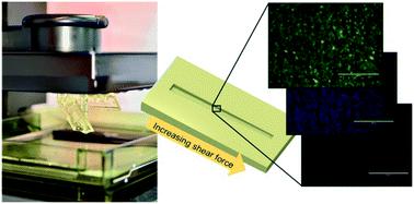
Three-dimensional (3D) printing has recently emerged as a cost-effective alternative for rapid prototyping of microfluidic devices. The feature resolution of stereolithography-based 3D printing is particularly well suited for manufacturing of continuous flow cell culture platforms. Poor cell adhesion or material-induced cell death may, however, limit the introduction of new materials to microfluidic cell culture. In this work, we characterized four commercially available materials commonly used in stereolithography-based 3D printing with respect to long-term (2 month) cell survival on native 3D printed surfaces. Cell proliferation rates, along with material-induced effects on apoptosis and cell survival, were examined in mouse embryonic fibroblasts. Additionally, the feasibility of Dental SG (material with the most favored properties) for culturing of human hepatocytes and human-induced pluripotent stem cells was evaluated. The strength of cell adhesion to Dental SG was further examined over a shear force gradient of 1–89 dyne per cm2 by using a custom-designed microfluidic shear force assay incorporating a 3D printed, tilted and tapered microchannel sealed with a polydimethylsiloxane lid. According to our results, autoclavation of the devices prior to cell seeding played the most important role in facilitating long-term cell survival on the native 3D printed surfaces with the shear force threshold in the range of 3–8 dyne per cm2.
中文翻译:

在微流体设备的立体光刻中使用的常见3D打印材料上的细胞粘附和增殖。
三维(3D)打印最近已经成为微流控设备快速原型制作的一种经济高效的替代方案。基于立体光刻的3D打印的功能分辨率特别适合于连续流动细胞培养平台的制造。然而,不良的细胞粘附或材料诱导的细胞死亡可能会限制将新材料引入微流细胞培养。在这项工作中,我们针对天然3D打印表面上的长期(2个月)细胞存活情况,对了四种通常用于基于立体光刻的3D打印中的市售材料进行了表征。在小鼠胚胎成纤维细胞中检查了细胞增殖率以及材料诱导的凋亡和细胞存活率影响。另外,评价了牙科SG(具有最有利特性的材料)用于培养人肝细胞和人诱导的多能干细胞的可行性。在1–89达因/厘米的剪切力梯度下,进一步检查了细胞对SG牙齿的粘附强度通过使用定制设计的微流体剪切力测定法(如图2所示),该测定法结合了用聚二甲基硅氧烷盖密封的3D打印,倾斜和锥形微通道。根据我们的结果,在植入细胞之前对设备进行高压灭菌在促进天然3D打印表面上的长期细胞存活方面发挥了最重要的作用,其剪切力阈值为3-8达因/ cm 2。
更新日期:2020-06-30
中文翻译:
在微流体设备的立体光刻中使用的常见3D打印材料上的细胞粘附和增殖。
三维(3D)打印最近已经成为微流控设备快速原型制作的一种经济高效的替代方案。基于立体光刻的3D打印的功能分辨率特别适合于连续流动细胞培养平台的制造。然而,不良的细胞粘附或材料诱导的细胞死亡可能会限制将新材料引入微流细胞培养。在这项工作中,我们针对天然3D打印表面上的长期(2个月)细胞存活情况,对了四种通常用于基于立体光刻的3D打印中的市售材料进行了表征。在小鼠胚胎成纤维细胞中检查了细胞增殖率以及材料诱导的凋亡和细胞存活率影响。另外,评价了牙科SG(具有最有利特性的材料)用于培养人肝细胞和人诱导的多能干细胞的可行性。在1–89达因/厘米的剪切力梯度下,进一步检查了细胞对SG牙齿的粘附强度通过使用定制设计的微流体剪切力测定法(如图2所示),该测定法结合了用聚二甲基硅氧烷盖密封的3D打印,倾斜和锥形微通道。根据我们的结果,在植入细胞之前对设备进行高压灭菌在促进天然3D打印表面上的长期细胞存活方面发挥了最重要的作用,其剪切力阈值为3-8达因/ cm 2。


 京公网安备 11010802027423号