当前位置:
X-MOL 学术
›
J. Cell. Physiol.
›
论文详情
Our official English website, www.x-mol.net, welcomes your
feedback! (Note: you will need to create a separate account there.)
The proinflammatory cytokines IL-1β and TNF-α modulate corneal epithelial wound healing through p16Ink4a suppressing STAT3 activity.
Journal of Cellular Physiology ( IF 4.5 ) Pub Date : 2020-05-31 , DOI: 10.1002/jcp.29823 Xiaolei Wang 1, 2 , Songmei Zhang 2 , Muchen Dong 3 , Yunqiu Li 4 , Qingjun Zhou 2 , Lingling Yang 2
Journal of Cellular Physiology ( IF 4.5 ) Pub Date : 2020-05-31 , DOI: 10.1002/jcp.29823 Xiaolei Wang 1, 2 , Songmei Zhang 2 , Muchen Dong 3 , Yunqiu Li 4 , Qingjun Zhou 2 , Lingling Yang 2
Affiliation
|
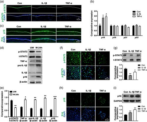
The proinflammatory cytokines interleukin‐1β (IL‐1β) and tumor necrosis factor‐α (TNF‐α) are involved in the corneal inflammatory response and wound healing following corneal injuries. However, the mechanism by which proinflammatory cytokines modulate corneal epithelial wound healing remains unclear. In this study, we found that IL‐1β or TNF‐α was transiently elevated during corneal epithelial wound healing in mice. After corneal epithelial debridement, persistent treatment with IL‐1β or TNF‐α restrained the level of phosphorylated signal transducer and activator of transcription 3 (p‐STAT3) and boosted the level of cell cycle inhibitor p16Ink4a, resulting in impaired corneal epithelial repair. When p16Ink4a was deleted, the p‐STAT3 level in corneal epithelium was enhanced and corneal epithelial wound healing was clearly accelerated. In diabetic mice, IL‐1β, TNF‐α, and p16Ink4a appeared a sustained and strong expression in the corneal epithelium, and p16Ink4a knockdown partially reverted the defective diabetic corneal epithelial repair. Furthermore, immunoprecipitation proved that p16Ink4a interacted with p‐STAT3 and thus possibly suppressed the STAT3 activity. Our findings revealed a novel mechanism that the proinflammatory cytokines modulate corneal epithelial wound healing via the p16Ink4a‐STAT3 signaling.
中文翻译:

促炎细胞因子IL-1β和TNF-α通过抑制STAT3活性的p16Ink4a调节角膜上皮伤口的愈合。
促炎细胞因子白介素-1β(IL-1β)和肿瘤坏死因子-α(TNF-α)参与角膜损伤后的角膜炎症反应和伤口愈合。但是,促炎细胞因子调节角膜上皮伤口愈合的机制仍不清楚。在这项研究中,我们发现小鼠角膜上皮伤口愈合期间IL-1β或TNF-α瞬时升高。角膜上皮清创后,持续用IL-1β或TNF-α治疗可抑制磷酸化信号转导子和转录激活因子3(p-STAT3)的水平,并提高细胞周期抑制剂p16 Ink4a的水平,从而导致角膜上皮修复受损。当p16 Ink4a去除角膜上皮中的p-STAT3水平,明显加速角膜上皮伤口愈合。在糖尿病小鼠中,IL-1β,TNF-α和p16 Ink4a在角膜上皮细胞中持续而强烈地表达,而p16 Ink4a的敲除可部分逆转糖尿病性角膜上皮的修复。此外,免疫沉淀证明p16 Ink4a与p-STAT3相互作用,因此可能抑制了STAT3活性。我们的发现揭示了一种新的机制,即促炎细胞因子通过p16 Ink4a- STAT3信号传导调节角膜上皮伤口愈合。
更新日期:2020-05-31
中文翻译:
促炎细胞因子IL-1β和TNF-α通过抑制STAT3活性的p16Ink4a调节角膜上皮伤口的愈合。
促炎细胞因子白介素-1β(IL-1β)和肿瘤坏死因子-α(TNF-α)参与角膜损伤后的角膜炎症反应和伤口愈合。但是,促炎细胞因子调节角膜上皮伤口愈合的机制仍不清楚。在这项研究中,我们发现小鼠角膜上皮伤口愈合期间IL-1β或TNF-α瞬时升高。角膜上皮清创后,持续用IL-1β或TNF-α治疗可抑制磷酸化信号转导子和转录激活因子3(p-STAT3)的水平,并提高细胞周期抑制剂p16 Ink4a的水平,从而导致角膜上皮修复受损。当p16 Ink4a去除角膜上皮中的p-STAT3水平,明显加速角膜上皮伤口愈合。在糖尿病小鼠中,IL-1β,TNF-α和p16 Ink4a在角膜上皮细胞中持续而强烈地表达,而p16 Ink4a的敲除可部分逆转糖尿病性角膜上皮的修复。此外,免疫沉淀证明p16 Ink4a与p-STAT3相互作用,因此可能抑制了STAT3活性。我们的发现揭示了一种新的机制,即促炎细胞因子通过p16 Ink4a- STAT3信号传导调节角膜上皮伤口愈合。


 京公网安备 11010802027423号