当前位置:
X-MOL 学术
›
J. Biophotonics
›
论文详情
Our official English website, www.x-mol.net, welcomes your
feedback! (Note: you will need to create a separate account there.)
Cell and nucleus refractive-index mapping by interferometric phase microscopy and rapid confocal fluorescence microscopy.
Journal of Biophotonics ( IF 2.0 ) Pub Date : 2020-07-06 , DOI: 10.1002/jbio.202000117 Shir Cohen-Maslaton 1 , Itay Barnea 1 , Almog Taieb 1 , Natan T Shaked 1
Journal of Biophotonics ( IF 2.0 ) Pub Date : 2020-07-06 , DOI: 10.1002/jbio.202000117 Shir Cohen-Maslaton 1 , Itay Barnea 1 , Almog Taieb 1 , Natan T Shaked 1
Affiliation
|
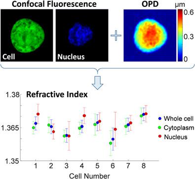
We present a multimodal technique for measuring the integral refractive index and the thickness of biological cells and their organelles by integrating interferometric phase microscopy (IPM) and rapid confocal fluorescence microscopy. First, the actual thickness maps of the cellular compartments are reconstructed using the confocal fluorescent sections, and then the optical path difference (OPD) map of the same cell is reconstructed using IPM. Based on the co‐registered data, the integral refractive index maps of the cell and its organelles are calculated. This technique enables rapidly measuring refractive index of live, dynamic cells, where IPM provides quantitative imaging capabilities and confocal fluorescence microscopy provides molecular specificity of the cell organelles. We acquire human colorectal adenocarcinoma cells and show that the integral refractive index values are similar for the whole cell, the cytoplasm and the nucleus on the population level, but significantly different on the single cell level.
中文翻译:

通过干涉相显微镜和快速共聚焦荧光显微镜对细胞和细胞核的折射率作图。
我们提出了一种通过整合干涉相显微镜(IPM)和快速共聚焦荧光显微镜来测量生物细胞及其细胞器的整体折射率和厚度的多峰技术。首先,使用共聚焦荧光部分重建细胞室的实际厚度图,然后使用IPM重建同一细胞的光程差(OPD)图。根据共同注册的数据,计算细胞及其细胞器的积分折射率图。这种技术能够快速测量活的,动态的细胞的折射率,其中IPM提供定量成像功能,而共聚焦荧光显微镜则提供细胞器的分子特异性。
更新日期:2020-07-06
中文翻译:
通过干涉相显微镜和快速共聚焦荧光显微镜对细胞和细胞核的折射率作图。
我们提出了一种通过整合干涉相显微镜(IPM)和快速共聚焦荧光显微镜来测量生物细胞及其细胞器的整体折射率和厚度的多峰技术。首先,使用共聚焦荧光部分重建细胞室的实际厚度图,然后使用IPM重建同一细胞的光程差(OPD)图。根据共同注册的数据,计算细胞及其细胞器的积分折射率图。这种技术能够快速测量活的,动态的细胞的折射率,其中IPM提供定量成像功能,而共聚焦荧光显微镜则提供细胞器的分子特异性。


 京公网安备 11010802027423号