Applied Materials Today ( IF 7.2 ) Pub Date : 2020-05-27 , DOI: 10.1016/j.apmt.2020.100706 Hoang Phuc Dang , Cedryck Vaquette , Tara Shabab , Román A. Pérez , Ying Yang , Tim R. Dargaville , Abbas Shafiee , Phong A. Tran
|
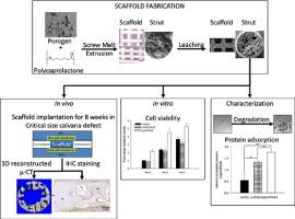
The application of 3D printed scaffolds for bone tissue regeneration has been explored in previous studies. In this study, we combined 3D printing with porogen leaching to develop scaffolds with dual-scale porosity and investigated their capability in guided bone regeneration in a rat critical size calvarial defect model. The scaffolds were additively manufactured from medical grade polycaprolactone (mPCL) doped with porogen microparticles having an average size of 22 μm, which were subsequently leached to create microscale porosity. Morphological analysis revealed an interconnected macroscale porosity of about 60% with an average pore size of 700 µm and intra-strut microscale pores with a porosity of nearly 40% and average pore size of 20–70 µm. The microscale porosity resulted in a 3-fold increase in the scaffolds’ surface area, a 2-fold enrichment in negatively charged surface groups, which did lead to significantly increased protein adsorption and faster hydrolysis-driven degradation in vitro. An in vitro blood clotting assay demonstrated an increased TGF-β1 release from the clots formed on the dual-scale porous scaffolds. In a rat calvarial defect, bone formation was found in both the macro- and microscale pores and was at a similar level when compared to calcium phosphate coated mPCL scaffolds.
中文翻译:
在大鼠颅骨缺损模型中用于引导骨再生的多孔3D打印支架
在先前的研究中已经探索了3D打印支架在骨组织再生中的应用。在这项研究中,我们结合了3D打印和致孔剂浸出技术来开发具有双尺度孔隙率的支架,并在大鼠关键尺寸颅盖骨缺损模型中研究了它们在引导骨再生中的能力。支架由掺杂有平均粒径为22μm的致孔剂微粒的医用级聚己内酯(mPCL)增材制造,随后进行沥滤以产生微孔。形态分析表明,相互关联的宏观孔隙度约为60%,平均孔径为700 µm,而杆内微尺度孔隙度为近40%,平均孔径为20-70 µm。微观孔隙率导致支架表面积增加了3倍,体外。一个体外血液凝血测定表明从形成在双尺度多孔支架的凝块增加的TGF-β1的释放。在大鼠颅盖骨缺损中,与磷酸钙包覆的mPCL支架相比,在大孔和小孔中均发现了骨形成,并且处于相似的水平。


 京公网安备 11010802027423号