当前位置:
X-MOL 学术
›
Spectrochim. Acta B. At. Spectrosc.
›
论文详情
Our official English website, www.x-mol.net, welcomes your
feedback! (Note: you will need to create a separate account there.)
Characterization of the elemental distribution of superalloy composite powders by micro beam X-ray fluorescence and laser-induced breakdown spectroscopy
Spectrochimica Acta Part B: Atomic Spectroscopy ( IF 3.2 ) Pub Date : 2020-07-01 , DOI: 10.1016/j.sab.2020.105896 Dong-ling Li , Zong-xin Liu , Lei Zhao , Xue-jing Shen , Hai-zhou Wang
Spectrochimica Acta Part B: Atomic Spectroscopy ( IF 3.2 ) Pub Date : 2020-07-01 , DOI: 10.1016/j.sab.2020.105896 Dong-ling Li , Zong-xin Liu , Lei Zhao , Xue-jing Shen , Hai-zhou Wang
|
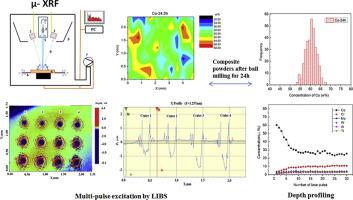
Abstract During the mechanical mixing of many types of powders, mechanical alloying will occur between powders of different components and particle sizes, accompanied by a series of complex physical and chemical changes. In this study, micro beam X-ray fluorescence (μ-XRF) and laser-induced breakdown spectroscopy (LIBS) were used to analyze the surface composition distribution and depth profile of cobalt and superalloy composite powders mixed for different ball-milling times. In addition, the variation of the content of the metal composite powders from the surface to the interior was determined. X-ray diffraction, scanning electron microscopy combined with energy-dispersive X-ray spectroscopy, and inductively coupled plasma atomic emission spectrometry were also used to characterize the morphology, structure, and the average content of the composite metal powders. The effects of the ball-milling time on the morphology, composition distribution, and microstructure of the composite metal powder were studied. Increasing ball-milling time led to improvement of the dispersion of cobalt powder in the superalloy composite. Fine cobalt particles were gradually attached to the surface of large superalloy particles by the mechanical alloying process. The cobalt contents determined by μ-XRF were clearly higher than the designed values owing to cobalt enrichment on the surface of the powder particles. Depth distribution analysis by LIBS also indicated the occurrence of cobalt segregation in the outer layer of composite particles with an enrichment thickness of normally less than 10 μm.
中文翻译:

用微束 X 射线荧光和激光诱导击穿光谱表征高温合金复合粉末的元素分布
摘要 多种粉体在机械混合过程中,不同成分和粒径的粉体之间会发生机械合金化,并伴随着一系列复杂的物理化学变化。在这项研究中,微束X射线荧光(μ-XRF)和激光诱导击穿光谱(LIBS)被用来分析不同球磨时间混合的钴和高温合金复合粉末的表面成分分布和深度分布。此外,测定了金属复合粉末的含量从表面到内部的变化。X射线衍射、扫描电子显微镜结合能量色散X射线光谱和电感耦合等离子体原子发射光谱也被用来表征形貌、结构、以及复合金属粉末的平均含量。研究了球磨时间对复合金属粉末形貌、成分分布和微观结构的影响。增加球磨时间导致钴粉在高温合金复合材料中的分散性改善。通过机械合金化过程,细小的钴颗粒逐渐附着在大的高温合金颗粒的表面。由于粉末颗粒表面的钴富集,μ-XRF 测定的钴含量明显高于设计值。LIBS 的深度分布分析也表明在复合颗粒的外层发生了钴偏析,富集厚度通常小于 10 μm。研究了复合金属粉末的成分分布和微观结构。增加球磨时间导致钴粉在高温合金复合材料中的分散性改善。通过机械合金化过程,细小的钴颗粒逐渐附着在大的高温合金颗粒的表面。由于粉末颗粒表面的钴富集,μ-XRF 测定的钴含量明显高于设计值。LIBS 的深度分布分析也表明在复合颗粒的外层发生了钴偏析,富集厚度通常小于 10 μm。研究了复合金属粉末的成分分布和微观结构。增加球磨时间导致钴粉在高温合金复合材料中的分散性改善。通过机械合金化过程,细小的钴颗粒逐渐附着在大的高温合金颗粒的表面。由于粉末颗粒表面的钴富集,μ-XRF 测定的钴含量明显高于设计值。LIBS 的深度分布分析也表明在复合颗粒的外层发生了钴偏析,富集厚度通常小于 10 μm。增加球磨时间导致钴粉在高温合金复合材料中的分散性改善。通过机械合金化过程,细小的钴颗粒逐渐附着在大的高温合金颗粒的表面。由于粉末颗粒表面的钴富集,μ-XRF 测定的钴含量明显高于设计值。LIBS 的深度分布分析也表明在复合颗粒的外层发生了钴偏析,富集厚度通常小于 10 μm。增加球磨时间导致钴粉在高温合金复合材料中的分散性改善。通过机械合金化过程,细小的钴颗粒逐渐附着在大的高温合金颗粒的表面。由于粉末颗粒表面的钴富集,μ-XRF 测定的钴含量明显高于设计值。LIBS 的深度分布分析也表明在复合颗粒的外层发生了钴偏析,富集厚度通常小于 10 μm。
更新日期:2020-07-01
中文翻译:
用微束 X 射线荧光和激光诱导击穿光谱表征高温合金复合粉末的元素分布
摘要 多种粉体在机械混合过程中,不同成分和粒径的粉体之间会发生机械合金化,并伴随着一系列复杂的物理化学变化。在这项研究中,微束X射线荧光(μ-XRF)和激光诱导击穿光谱(LIBS)被用来分析不同球磨时间混合的钴和高温合金复合粉末的表面成分分布和深度分布。此外,测定了金属复合粉末的含量从表面到内部的变化。X射线衍射、扫描电子显微镜结合能量色散X射线光谱和电感耦合等离子体原子发射光谱也被用来表征形貌、结构、以及复合金属粉末的平均含量。研究了球磨时间对复合金属粉末形貌、成分分布和微观结构的影响。增加球磨时间导致钴粉在高温合金复合材料中的分散性改善。通过机械合金化过程,细小的钴颗粒逐渐附着在大的高温合金颗粒的表面。由于粉末颗粒表面的钴富集,μ-XRF 测定的钴含量明显高于设计值。LIBS 的深度分布分析也表明在复合颗粒的外层发生了钴偏析,富集厚度通常小于 10 μm。研究了复合金属粉末的成分分布和微观结构。增加球磨时间导致钴粉在高温合金复合材料中的分散性改善。通过机械合金化过程,细小的钴颗粒逐渐附着在大的高温合金颗粒的表面。由于粉末颗粒表面的钴富集,μ-XRF 测定的钴含量明显高于设计值。LIBS 的深度分布分析也表明在复合颗粒的外层发生了钴偏析,富集厚度通常小于 10 μm。研究了复合金属粉末的成分分布和微观结构。增加球磨时间导致钴粉在高温合金复合材料中的分散性改善。通过机械合金化过程,细小的钴颗粒逐渐附着在大的高温合金颗粒的表面。由于粉末颗粒表面的钴富集,μ-XRF 测定的钴含量明显高于设计值。LIBS 的深度分布分析也表明在复合颗粒的外层发生了钴偏析,富集厚度通常小于 10 μm。增加球磨时间导致钴粉在高温合金复合材料中的分散性改善。通过机械合金化过程,细小的钴颗粒逐渐附着在大的高温合金颗粒的表面。由于粉末颗粒表面的钴富集,μ-XRF 测定的钴含量明显高于设计值。LIBS 的深度分布分析也表明在复合颗粒的外层发生了钴偏析,富集厚度通常小于 10 μm。增加球磨时间导致钴粉在高温合金复合材料中的分散性改善。通过机械合金化过程,细小的钴颗粒逐渐附着在大的高温合金颗粒的表面。由于粉末颗粒表面的钴富集,μ-XRF 测定的钴含量明显高于设计值。LIBS 的深度分布分析也表明在复合颗粒的外层发生了钴偏析,富集厚度通常小于 10 μm。


 京公网安备 11010802027423号