Respiratory Medicine ( IF 3.5 ) Pub Date : 2020-05-25 , DOI: 10.1016/j.rmed.2020.106043 Karina Loor 1 , Esther Pallisa 2 , Mario Culebras 3 , Maria Deu 4 , Antonio Álvarez 3 , Irene Sansano 5 , Cristina Berastegui 3 , David Clofent 1 , Eva Polverino 6 , Javier de Gracia 6
|
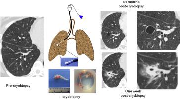
Background
The occurrence of radiological opacities post-transbronchial cryobiopsy may pose serious difficulties in differential diagnosis and management of lung allografts. This prospective study evaluated the frequency, characteristics, and evolution of new lung opacities after performing transbronchial cryobiopsy.
Methods
From February 2018 to June 2018, 22 of 51 consecutive patients with an indication for transbronchial cryobiopsy underwent computed tomography (CT) of the thorax before and at 1, 4, and 8 weeks post-cryobiopsy. New CT images, required by the transplant team, were also evaluated during the next 6 months. Histological findings of transbronchial cryobiopsy and microbiological studies on bronchoalveolar lavage were evaluated as risk factors for opacities.
Results
After obtaining 112 cryobiopsy samples, 46 opacities >10 mm, including ground-glass, solid, cavitated, or a combination of these lesions were observed in 20 (91%) patients on post-cryobiopsy CT. All ground-glasses opacities on CT disappeared at 4 weeks. A single cavitated opacity persisted at 6 months. The remaining opacities disappeared or were decreased to <10 mm by 8 weeks. No correlations of the number, type, or evolution of opacities with the number or volume of cryobiopsy samples obtained, or with the histological diagnosis, type of transplant, or microbiologic culture results were observed.
Conclusion
New pulmonary opacities >10 mm occur frequently after transbronchial cryobiopsy; a few may persist beyond 6 months. CT studies are recommended before implementing transbronchial cryobiopsy, whenever possible.
中文翻译:
经支气管冷冻活检后肺同种异体移植物出现新的混浊。
背景
经支气管冷冻活检后放射学混浊的发生可能对肺同种异体移植物的鉴别诊断和管理造成严重困难。这项前瞻性研究评估了经支气管冷冻活检后新肺部混浊的频率、特征和演变。
方法
从 2018 年 2 月到 2018 年 6 月,连续 51 名有经支气管冷冻活检指征的患者中有 22 名在冷冻活检前和冷冻活检后 1、4 和 8 周时接受了胸部计算机断层扫描 (CT)。移植团队需要的新 CT 图像也在接下来的 6 个月内进行了评估。经支气管冷冻活检的组织学发现和支气管肺泡灌洗的微生物学研究被评估为混浊的危险因素。
结果
在获得 112 个冷冻活检样本后,在 20 名 (91%) 患者的冷冻活检后 CT 中观察到 46 个 >10 mm 的混浊,包括毛玻璃样、实体、空洞或这些病变的组合。CT 上的所有毛玻璃混浊在 4 周时消失。单个空洞混浊在 6 个月时持续存在。到 8 周时,剩余的混浊消失或减少到 <10 毫米。未观察到混浊的数量、类型或演变与获得的冷冻活检样本的数量或体积,或与组织学诊断、移植类型或微生物培养结果的相关性。
结论
经支气管冷冻活检后常出现>10mm的新肺部混浊;少数可能会持续超过 6 个月。只要有可能,建议在实施经支气管冷冻活检之前进行 CT 检查。


 京公网安备 11010802027423号