当前位置:
X-MOL 学术
›
J. Comp. Neurol.
›
论文详情
Our official English website, www.x-mol.net, welcomes your
feedback! (Note: you will need to create a separate account there.)
Distribution and localization of phosphatidylinositol 5-phosphate, 4-kinase alpha and beta in the brain.
The Journal of Comparative Neurology ( IF 2.3 ) Pub Date : 2020-05-24 , DOI: 10.1002/cne.24956 Evan K Noch 1, 2 , Isaiah Yim 1 , Teresa A Milner 3, 4 , Lewis C Cantley 1
The Journal of Comparative Neurology ( IF 2.3 ) Pub Date : 2020-05-24 , DOI: 10.1002/cne.24956 Evan K Noch 1, 2 , Isaiah Yim 1 , Teresa A Milner 3, 4 , Lewis C Cantley 1
Affiliation
|
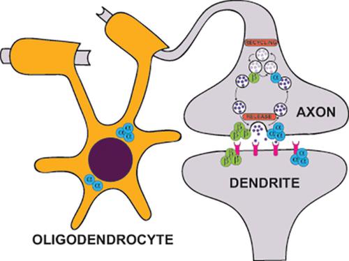
Phosphatidylinositol‐4,5‐bisphosphate (PI‐4,5‐P2) is critical for synaptic vesicle docking and fusion and generation of the second messengers, diacylglycerol and inositol‐1,4,5‐trisphosphate. PI‐4,5‐P2 can be generated by two families of kinases: type 1 phosphatidylinositol‐4‐phosphate 5‐kinases, encoded by PIP5K1A, PIP5K1B and PIP5K1C, and type 2 phosphatidylinositol‐5‐phosphate 4‐kinases, encoded by PIP4K2A, PIP4K2B, and PIP4K2C. While the roles of the type 1 enzymes in brain function have been extensively studied, the roles of the type 2 enzymes are poorly understood. Using selective antibodies validated by genetic deletion of pip4k2a or pip4k2b in mouse brain, we characterized the location of the enzymes, PI5P4Kα and PI5P4Kβ, encoded by these genes. In mice, we demonstrate that PI5P4Kα is expressed in adulthood, whereas PI5P4Kβ is expressed early in development. PI5P4Kα localizes to white matter tracts, especially the corpus callosum, and at a low level in neurons, while PI5P4Kβ is expressed in neuronal populations, especially hippocampus and cortex. Dual labeling studies demonstrate that PI5P4Kα co‐localizes with the oligodendrocyte marker, Olig2, whereas PI5P4Kβ co‐localizes with the neuronal marker, NeuN. Ultrastructural analysis demonstrates that both kinases are contained in axon terminals and dendritic spines adjacent to the synaptic membrane, which support a potential role in synaptic transmission. Immunoperoxidase analysis of macaque and human brain tissue demonstrate a conserved pattern for PI5P4Kα and PI5P4Kβ. These results highlight the diverse cell‐autonomous expression of PI5P4Kα and PI5P4Kβ and support further exploration into their role in synaptic function in the brain.
中文翻译:

磷脂酰肌醇 5-磷酸、4-激酶 α 和 β 在脑中的分布和定位。
磷脂酰肌醇-4,5-二磷酸 (PI-4,5-P 2 ) 对于突触小泡对接和融合以及第二信使二酰基甘油和肌醇-1,4,5-三磷酸的产生至关重要。PI-4,5-P 2可由两个激酶家族产生:1 型磷脂酰肌醇-4-磷酸 5-激酶,由 PIP5K1A、PIP5K1B 和 PIP5K1C 编码,2 型磷脂酰肌醇-5-磷酸 4-激酶,由 PIP4K2A、PIP4K2B 和 PIP4K2C 编码。虽然 1 型酶在脑功能中的作用已被广泛研究,但 2 型酶的作用却知之甚少。使用通过小鼠脑中 pip4k2a 或 pip4k2b 基因缺失验证的选择性抗体,我们表征了由这些基因编码的酶 PI5P4Kα 和 PI5P4Kβ 的位置。在小鼠中,我们证明 PI5P4Kα 在成年期表达,而 PI5P4Kβ 在发育早期表达。PI5P4Kα 定位于白质束,尤其是胼胝体,在神经元中水平较低,而 PI5P4Kβ 在神经元群中表达,尤其是海马体和皮层。双标记研究表明,PI5P4Kα 与少突胶质细胞标记 Olig2 共定位,而 PI5P4Kβ 与神经元标记 NeuN 共定位。超微结构分析表明,这两种激酶都包含在与突触膜相邻的轴突末端和树突棘中,这支持了突触传递中的潜在作用。猕猴和人脑组织的免疫过氧化物酶分析证明了 PI5P4Kα 和 PI5P4Kβ 的保守模式。这些结果突出了 PI5P4Kα 和 PI5P4Kβ 的不同细胞自主表达,并支持进一步探索它们在大脑突触功能中的作用。超微结构分析表明,这两种激酶都包含在与突触膜相邻的轴突末端和树突棘中,这支持了突触传递中的潜在作用。猕猴和人脑组织的免疫过氧化物酶分析证明了 PI5P4Kα 和 PI5P4Kβ 的保守模式。这些结果突出了 PI5P4Kα 和 PI5P4Kβ 的不同细胞自主表达,并支持进一步探索它们在大脑突触功能中的作用。超微结构分析表明,这两种激酶都包含在与突触膜相邻的轴突末端和树突棘中,这支持了突触传递中的潜在作用。猕猴和人脑组织的免疫过氧化物酶分析证明了 PI5P4Kα 和 PI5P4Kβ 的保守模式。这些结果突出了 PI5P4Kα 和 PI5P4Kβ 的不同细胞自主表达,并支持进一步探索它们在大脑突触功能中的作用。
更新日期:2020-06-27
中文翻译:
磷脂酰肌醇 5-磷酸、4-激酶 α 和 β 在脑中的分布和定位。
磷脂酰肌醇-4,5-二磷酸 (PI-4,5-P 2 ) 对于突触小泡对接和融合以及第二信使二酰基甘油和肌醇-1,4,5-三磷酸的产生至关重要。PI-4,5-P 2可由两个激酶家族产生:1 型磷脂酰肌醇-4-磷酸 5-激酶,由 PIP5K1A、PIP5K1B 和 PIP5K1C 编码,2 型磷脂酰肌醇-5-磷酸 4-激酶,由 PIP4K2A、PIP4K2B 和 PIP4K2C 编码。虽然 1 型酶在脑功能中的作用已被广泛研究,但 2 型酶的作用却知之甚少。使用通过小鼠脑中 pip4k2a 或 pip4k2b 基因缺失验证的选择性抗体,我们表征了由这些基因编码的酶 PI5P4Kα 和 PI5P4Kβ 的位置。在小鼠中,我们证明 PI5P4Kα 在成年期表达,而 PI5P4Kβ 在发育早期表达。PI5P4Kα 定位于白质束,尤其是胼胝体,在神经元中水平较低,而 PI5P4Kβ 在神经元群中表达,尤其是海马体和皮层。双标记研究表明,PI5P4Kα 与少突胶质细胞标记 Olig2 共定位,而 PI5P4Kβ 与神经元标记 NeuN 共定位。超微结构分析表明,这两种激酶都包含在与突触膜相邻的轴突末端和树突棘中,这支持了突触传递中的潜在作用。猕猴和人脑组织的免疫过氧化物酶分析证明了 PI5P4Kα 和 PI5P4Kβ 的保守模式。这些结果突出了 PI5P4Kα 和 PI5P4Kβ 的不同细胞自主表达,并支持进一步探索它们在大脑突触功能中的作用。超微结构分析表明,这两种激酶都包含在与突触膜相邻的轴突末端和树突棘中,这支持了突触传递中的潜在作用。猕猴和人脑组织的免疫过氧化物酶分析证明了 PI5P4Kα 和 PI5P4Kβ 的保守模式。这些结果突出了 PI5P4Kα 和 PI5P4Kβ 的不同细胞自主表达,并支持进一步探索它们在大脑突触功能中的作用。超微结构分析表明,这两种激酶都包含在与突触膜相邻的轴突末端和树突棘中,这支持了突触传递中的潜在作用。猕猴和人脑组织的免疫过氧化物酶分析证明了 PI5P4Kα 和 PI5P4Kβ 的保守模式。这些结果突出了 PI5P4Kα 和 PI5P4Kβ 的不同细胞自主表达,并支持进一步探索它们在大脑突触功能中的作用。


 京公网安备 11010802027423号