当前位置:
X-MOL 学术
›
Cytom. Part A
›
论文详情
Our official English website, www.x-mol.net, welcomes your
feedback! (Note: you will need to create a separate account there.)
Phenotypic Analysis of the Mouse Hematopoietic Hierarchy Using Spectral Cytometry: From Stem Cell Subsets to Early Progenitor Compartments.
Cytometry Part A ( IF 2.5 ) Pub Date : 2020-05-25 , DOI: 10.1002/cyto.a.24041 Michael Solomon 1 , Monica DeLay 2 , Damien Reynaud 1, 3
Cytometry Part A ( IF 2.5 ) Pub Date : 2020-05-25 , DOI: 10.1002/cyto.a.24041 Michael Solomon 1 , Monica DeLay 2 , Damien Reynaud 1, 3
Affiliation
|
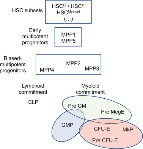
Phenotypic analysis by flow cytometry is one of the most utilized primary tools to study the hematopoietic system. Here, we present a complex panel designed for spectral flow cytometry that allows for the in‐depth analysis of the mouse hematopoietic stem and progenitor compartments. The developed panel encompasses the hematopoietic stem cell (HSC) compartment, an array of multipotent progenitors with early marks of lineage specification and a series of progenitors associated with lymphoid, granulo‐macrophagic, megakaryocytic and erythroid lineage commitment. It has a built‐in redundancy for key markers known to decipher the fine architecture of the HSC compartment by segregating subsets with different functional potential. As a resource, we used this panel to provide a snapshot view of the evolution of these phenotypically defined hematopoietic compartments during the life of the animals. We show that by using a spectral cytometer, this panel is compatible with the analysis of GFP‐expressing gene‐reporter mice across the hematopoietic system. We leverage this tool to determine how previously described markers such as CD150, CD34, CD105, CD41, ECPR, and CD49b define specific HSC subsets and confirm that high expression of the transcription factor Gfi1 is a hallmark of the most primitive HSC compartment. Altogether, our results provide a convenient protocol to obtain in one analysis a more extensive view of the hematopoietic architecture in mouse models. Our results could also serve as a base for further development of high‐end panels leveraging spectral flow cytometry beyond the 15‐fluorochrome panel presented in this report. © 2020 International Society for Advancement of Cytometry
中文翻译:

使用光谱细胞计数法对小鼠造血层级进行表型分析:从干细胞亚群到早期祖细胞室。
流式细胞术表型分析是研究造血系统最常用的主要工具之一。在这里,我们展示了一个为光谱流式细胞术设计的复杂面板,可以对小鼠造血干细胞和祖细胞进行深入分析。开发的面板包括造血干细胞 (HSC) 区室、一系列具有早期谱系规范标记的多能祖细胞,以及一系列与淋巴、粒-巨噬细胞、巨核细胞和红细胞谱系相关的祖细胞。它对已知的关键标记具有内置冗余,通过分离具有不同功能潜力的子集来破译 HSC 区室的精细结构。作为资源,我们使用该面板提供了这些表型定义的造血区室在动物生命期间进化的快照视图。我们表明,通过使用光谱细胞仪,该面板与整个造血系统中表达 GFP 的基因报告小鼠的分析兼容。我们利用此工具来确定先前描述的标记(如 CD150、CD34、CD105、CD41、ECPR 和 CD49b)如何定义特定的 HSC 子集,并确认转录因子 Gfi1 的高表达是最原始 HSC 区室的标志。总而言之,我们的结果提供了一种方便的协议,可以在一次分析中获得更广泛的小鼠模型造血结构视图。我们的结果还可以作为进一步开发利用光谱流式细胞术的高端面板的基础,而不是本报告中介绍的 15 荧光染料面板。© 2020 国际细胞计量学促进会
更新日期:2020-05-25
中文翻译:
使用光谱细胞计数法对小鼠造血层级进行表型分析:从干细胞亚群到早期祖细胞室。
流式细胞术表型分析是研究造血系统最常用的主要工具之一。在这里,我们展示了一个为光谱流式细胞术设计的复杂面板,可以对小鼠造血干细胞和祖细胞进行深入分析。开发的面板包括造血干细胞 (HSC) 区室、一系列具有早期谱系规范标记的多能祖细胞,以及一系列与淋巴、粒-巨噬细胞、巨核细胞和红细胞谱系相关的祖细胞。它对已知的关键标记具有内置冗余,通过分离具有不同功能潜力的子集来破译 HSC 区室的精细结构。作为资源,我们使用该面板提供了这些表型定义的造血区室在动物生命期间进化的快照视图。我们表明,通过使用光谱细胞仪,该面板与整个造血系统中表达 GFP 的基因报告小鼠的分析兼容。我们利用此工具来确定先前描述的标记(如 CD150、CD34、CD105、CD41、ECPR 和 CD49b)如何定义特定的 HSC 子集,并确认转录因子 Gfi1 的高表达是最原始 HSC 区室的标志。总而言之,我们的结果提供了一种方便的协议,可以在一次分析中获得更广泛的小鼠模型造血结构视图。我们的结果还可以作为进一步开发利用光谱流式细胞术的高端面板的基础,而不是本报告中介绍的 15 荧光染料面板。© 2020 国际细胞计量学促进会


 京公网安备 11010802027423号