当前位置:
X-MOL 学术
›
J. Biophotonics
›
论文详情
Our official English website, www.x-mol.net, welcomes your
feedback! (Note: you will need to create a separate account there.)
3D morphological and biophysical changes in a single tachyzoite and its infected cells using three-dimensional quantitative phase imaging.
Journal of Biophotonics ( IF 2.0 ) Pub Date : 2020-06-05 , DOI: 10.1002/jbio.202000055 Egy Rahman Firdaus 1 , Ji-Hoon Park 1 , Seong-Kyun Lee 1 , YongKeun Park 2 , Guang-Ho Cha 3 , Eun-Taek Han 1
Journal of Biophotonics ( IF 2.0 ) Pub Date : 2020-06-05 , DOI: 10.1002/jbio.202000055 Egy Rahman Firdaus 1 , Ji-Hoon Park 1 , Seong-Kyun Lee 1 , YongKeun Park 2 , Guang-Ho Cha 3 , Eun-Taek Han 1
Affiliation
|
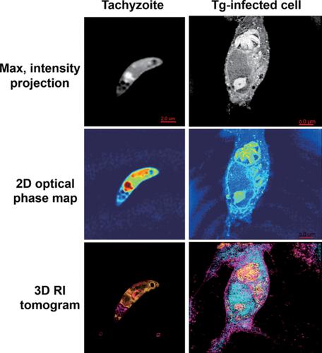
Toxoplasma gondii is an apicomplexan parasite that causes toxoplasmosis in the human body and commonly infects warm‐blooded organisms. Pathophysiology of its diseases is still an interesting issue to be studied since T gondii can infect nearly all nucleated cells. Imaging techniques are crucial for studying its pathophysiology. In T gondii‐infected cells structural and biochemical alterations occurred. To study that modification, we use digital holotomography to investigate the structure and biochemical alteration of single tachyzoite and its infected cells in a label‐free and quantitative manner. Quantification analysis was done by measuring the refractive index distribution, which provides information about the concentration and dry mass of individual cells. This study showed that holotomography could be effectively used to identify the structural and biochemical alteration in tremendously different cells in supporting pathophysiological research in particular for T gondii‐caused diseases.
中文翻译:

使用三维定量相位成像,单个速殖子及其感染的细胞中的3D形态和生物物理变化。
弓形虫是一种寄生虫,会引起人的弓形虫病,并通常感染温血生物。由于弓形虫可感染几乎所有有核细胞,因此其疾病的病理生理学仍是一个值得研究的有趣问题。成像技术对于研究其病理生理至关重要。在贡迪被感染细胞发生结构和生化改变。为了研究这种修饰,我们使用数字全息术以无标记和定量的方式研究了单个速殖子及其感染细胞的结构和生化变化。通过测量折射率分布进行定量分析,该折射率分布提供有关单个细胞的浓度和干质量的信息。这项研究表明,全息照相法可有效地用于识别极为不同的细胞中的结构和生化改变,从而支持病理生理学研究,尤其是针对弓形虫引起的疾病。
更新日期:2020-06-05
中文翻译:
使用三维定量相位成像,单个速殖子及其感染的细胞中的3D形态和生物物理变化。
弓形虫是一种寄生虫,会引起人的弓形虫病,并通常感染温血生物。由于弓形虫可感染几乎所有有核细胞,因此其疾病的病理生理学仍是一个值得研究的有趣问题。成像技术对于研究其病理生理至关重要。在贡迪被感染细胞发生结构和生化改变。为了研究这种修饰,我们使用数字全息术以无标记和定量的方式研究了单个速殖子及其感染细胞的结构和生化变化。通过测量折射率分布进行定量分析,该折射率分布提供有关单个细胞的浓度和干质量的信息。这项研究表明,全息照相法可有效地用于识别极为不同的细胞中的结构和生化改变,从而支持病理生理学研究,尤其是针对弓形虫引起的疾病。


 京公网安备 11010802027423号