Journal of Neuroscience Methods ( IF 2.7 ) Pub Date : 2020-05-22 , DOI: 10.1016/j.jneumeth.2020.108792 Jared Leichner 1 , Wei-Chiang Lin 1
|
Background
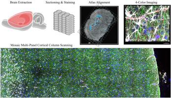
Immunofluorescent staining coupled with axial optical sectioning allows for assessment of native three-dimensional structure of brain tissue. Typical challenges of analyzing network structure include limitations driven by magnification/field of view, spatial resolution, tissue thickness, staining quality of dense cell types, data quantifiability and the quantity of simultaneous staining targets.
New Method
This manuscript demonstrates many methodological advancements. Software-aided alignment of the cortical slice and stereotaxic atlas maximizes ROI-identification accuracy. Tissue compression during antigen retrieval enhances epitope availability without damaging tissue. A thorough factorial experiment focusing on Smi-311 staining highlights the enhancements in image quality from our extended staining protocol. Mosaic scanning techniques and subsequent four-channel alignment ensures high data quality.
Results
Cortical column datasets [800μm x 3000μm x 70μm] utilizing sequential optical sectioning were successfully generated from three rats. Each rat provided three coronal sections in each of two regions, M1 and S1BF, from which data cubes were generated per hemisphere, totaling 36 high-magnification four-color datasets.
Comparison with Existing Method(s)
Typical confocal assessments of brain tissue do not utilize such thick tissue slices nor collect entire cortical columns from the cortical surface to the grey/white interface at a resolution that can map fine filamentous processes. The simultaneous collection of our four specific structural markers - neuronal, astrocytic, vascular and nuclear – is novel and the quantitative optimization of staining protocols through a factorial design rare.
Conclusions
Building upon this preliminary success in protocol development, future work will encompass volumetric modeling and quantitative analysis of regional network architecture.
中文翻译:
通过大鼠皮层柱对4种荧光成分进行成像和分析的进展。
背景
免疫荧光染色与轴向光学切片相结合,可以评估脑组织的天然三维结构。分析网络结构的典型挑战包括由放大率/视野,空间分辨率,组织厚度,致密细胞类型的染色质量,数据可量化性和同时染色目标的数量所驱动的局限性。
新方法
该手稿展示了许多方法上的进步。皮质切片和立体定位图谱的软件辅助对齐可最大程度地提高ROI识别的准确性。抗原修复过程中的组织压缩可提高表位的利用率,而不会损坏组织。一项针对Smi-311染色的彻底析因实验强调了我们扩展的染色方案可提高图像质量。马赛克扫描技术和随后的四通道对齐确保了高数据质量。
结果
从三只大鼠中成功地利用连续光学切片获得了皮质柱数据集[800μmx3000μmx70μm]。每只大鼠在M1和S1BF两个区域中的每个区域提供了三个冠状切片,从每个半球生成了数据立方体,总共形成了36个高放大率的四色数据集。
与现有方法的比较
对脑组织的典型共聚焦评估既不利用这样厚的组织切片,也不以可绘制精细的丝状过程的分辨率收集从皮质表面到灰色/白色界面的整个皮质柱。同时收集我们四个特定的结构标记-神经元,星形细胞,血管和核-是新颖的,并且通过因子设计对染色方案进行定量优化的情况很少。
结论
基于协议开发的初步成功,未来的工作将包括区域网络体系结构的体积建模和定量分析。


 京公网安备 11010802027423号