Computer Methods and Programs in Biomedicine ( IF 4.9 ) Pub Date : 2020-05-22 , DOI: 10.1016/j.cmpb.2020.105533 Yang Li 1 , Yu-Qian Zhao 2 , Fan Zhang 3 , Miao Liao 4 , Ling-Li Yu 3 , Bai-Fan Chen 5 , Yan-Jin Wang 6
|
Background and objective
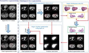
Liver segmentation from abdominal CT volumes is a primary step for computer-aided surgery and liver disease diagnosis. However, accurate liver segmentation remains a challenging task for intensity inhomogeneity and serious pathologies occurring in liver CT volume. This paper presents a novel framework for accurate liver segmentation from CT images.
Methods
Firstly, a novel level set integrated with intensity bias and position constraint is applied, and for normal liver, the generated liver regions are regarded as the final results. Then, for pathological liver, a sparse shape composition (SSC)-based method is presented to refine liver shapes, followed by an improved graph cut to further optimize segmentation results. The level set-based method is capable of overcoming intensity inhomogeneity in object regions, and the SSC- and graph cut-based strategy has outstanding power to address under-segmentation appearing in pathological livers.
Results
The experiments conducted on public databases SLIVER07 and 3Dircadb show that the proposed method can segment both healthy and pathological liver effectively. The segmentation performance in terms of mean ASD, RMSD, MSD, VOE and RVD on SLIVER07 are 0.9mm, 1.8mm, 19.4mm, 5.1% and 0.1%, respectively, and on 3Dircadb are 1.6mm, 3.1mm, 27.2mm, 9.2% and 0.5%, respectively, which outperforms many existing methods.
Conclusions
The proposed method does not require complex training procedure on numerous liver samples, and has satisfying and robust segmentation performance on both normal and pathological liver in various shapes.
中文翻译:
根据水平集和稀疏形状组成从腹部CT量进行肝脏分割。
背景和目标
从腹部CT量进行肝脏分割是计算机辅助手术和肝病诊断的第一步。然而,对于肝脏CT容积中出现的强度不均匀和严重病理,准确的肝脏分割仍然是一项艰巨的任务。本文提出了一种从CT图像准确分割肝脏的新颖框架。
方法
首先,应用结合强度偏差和位置约束的新型水平集,对于正常肝脏,将生成的肝脏区域视为最终结果。然后,对于病理性肝脏,提出了一种基于稀疏形状成分(SSC)的方法来细化肝脏形状,然后进行改进的图形切割以进一步优化分割结果。基于水平集的方法能够克服对象区域中的强度不均匀性,并且基于SSC和图割的策略具有出色的能力来应对病理肝脏中出现的节段不足。
结果
在公共数据库SLIVER07和3Dircadb上进行的实验表明,该方法可以有效地分割健康肝脏和病理肝脏。SLIVER07的平均ASD,RMSD,MSD,VOE和RVD分割性能分别为0.9mm,1.8mm,19.4mm,5.1%和0.1%,而3Dircadb的分割性能分别为1.6mm,3.1mm,27.2mm,9.2 %和0.5%,分别优于许多现有方法。
结论
所提出的方法不需要对大量肝脏样品进行复杂的训练过程,并且在各种形状的正常肝脏和病理肝脏上均具有令人满意的鲁棒分割性能。


 京公网安备 11010802027423号