当前位置:
X-MOL 学术
›
J. Chem. Neuroanat.
›
论文详情
Our official English website, www.x-mol.net, welcomes your feedback! (Note: you will need to create a separate account there.)
LC3 and ATG5 Overexpression and Neuronal Cell Death in the Prefrontal Cortex of Postmortem Chronic Methamphetamine Users
Journal of Chemical Neuroanatomy ( IF 2.8 ) Pub Date : 2020-09-01 , DOI: 10.1016/j.jchemneu.2020.101802 Shahrokh Khoshsirat 1 , Maryam Sadat Khoramgah 2 , Gholam-Reza Mahmoudiasl 3 , Mostafa Rezaei-Tavirani 4 , Mohammad-Amin Abdollahifar 5 , Foozhan Tahmasebinia 6 , Shahram Darabi 7 , Somayeh Niknazar 1 , Hojjat Allah Abbaszadeh 2
Journal of Chemical Neuroanatomy ( IF 2.8 ) Pub Date : 2020-09-01 , DOI: 10.1016/j.jchemneu.2020.101802 Shahrokh Khoshsirat 1 , Maryam Sadat Khoramgah 2 , Gholam-Reza Mahmoudiasl 3 , Mostafa Rezaei-Tavirani 4 , Mohammad-Amin Abdollahifar 5 , Foozhan Tahmasebinia 6 , Shahram Darabi 7 , Somayeh Niknazar 1 , Hojjat Allah Abbaszadeh 2
Affiliation
|
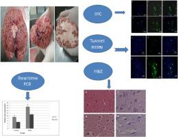
Methamphetamine (METH) abuse is accompanied by oxidative stress, METH-induced neurotoxicity, and apoptosis. Oxidative stress has devastating effects on the structure of proteins and cells. Autophagy is an evolutionarily conserved intracellular regulated mechanism for orderly degradation of dysfunctional proteins or removing damaged organelles. The precise role of autophagy in oxidative stress-induced apoptosis of dopaminergic neuronal cells caused by METH has not clarified completely. In this study, we sought to evaluate the effects of METH abuse on autophagy in the prefrontal cortex of postmortem users, mainly focusing on the ATG5 and LC3 during neuroinflammation. Postmortem molecular and histological examination was done for two groups containing 12 non-addicted and 14 METH addicted cases. ATG5 and LC3 expression were analyzed by real-time PCR and immunohistochemistry (IHC) methods. Histopathological analysis was performed by stereological cell counting of neuronal cells using Hematoxylin and Eosin (H & E) staining technique. In order to detect DNA damage in the prefrontal lobe, Tunnel staining was performed. Real-time PCR and IHC assay showed overexpression of ATG5 and LC3 protein in the prefrontal cortex of Meth users. The cell death and neuronal degeneration were increased significantly based on Tunel assay and the stereological analysis in the Prefrontal cortex. Chronic METH exposure probably induces ATG5 and LC3 overexpression and neuronal cell death in the Prefrontal cortex of the postmortem cases.
中文翻译:

死后慢性甲基苯丙胺使用者前额叶皮层中的 LC3 和 ATG5 过度表达和神经元细胞死亡
甲基苯丙胺 (METH) 滥用伴随着氧化应激、METH 诱导的神经毒性和细胞凋亡。氧化应激对蛋白质和细胞的结构具有破坏性影响。自噬是一种进化上保守的细胞内调节机制,用于有序降解功能失调的蛋白质或去除受损的细胞器。自噬在氧化应激诱导的 METH 引起的多巴胺能神经元细胞凋亡中的确切作用尚未完全阐明。在这项研究中,我们试图评估 METH 滥用对死后使用者前额叶皮层自噬的影响,主要关注神经炎症期间的 ATG5 和 LC3。对包含 12 个非成瘾病例和 14 个 METH 成瘾病例的两组进行了死后分子和组织学检查。通过实时 PCR 和免疫组织化学 (IHC) 方法分析 ATG5 和 LC3 的表达。使用苏木精和伊红 (H&E) 染色技术通过神经元细胞的立体细胞计数进行组织病理学分析。为了检测前额叶中的 DNA 损伤,进行了隧道染色。实时 PCR 和 IHC 分析显示,在 Meth 使用者的前额叶皮层中 ATG5 和 LC3 蛋白过度表达。根据 Tunel 测定和前额叶皮层的立体学分析,细胞死亡和神经元变性显着增加。慢性 METH 暴露可能会诱导死后病例的前额叶皮层中的 ATG5 和 LC3 过表达和神经元细胞死亡。使用苏木精和伊红 (H & E) 染色技术通过神经元细胞的立体细胞计数进行组织病理学分析。为了检测前额叶中的 DNA 损伤,进行了隧道染色。实时 PCR 和 IHC 分析显示,在 Meth 使用者的前额叶皮层中 ATG5 和 LC3 蛋白过度表达。根据 Tunel 测定和前额叶皮层的立体学分析,细胞死亡和神经元变性显着增加。慢性 METH 暴露可能会诱导死后病例的前额叶皮层中的 ATG5 和 LC3 过表达和神经元细胞死亡。使用苏木精和伊红 (H&E) 染色技术通过神经元细胞的立体细胞计数进行组织病理学分析。为了检测前额叶中的 DNA 损伤,进行了隧道染色。实时 PCR 和 IHC 分析显示,在 Meth 使用者的前额叶皮层中 ATG5 和 LC3 蛋白过度表达。根据 Tunel 测定和前额叶皮层的立体学分析,细胞死亡和神经元变性显着增加。慢性 METH 暴露可能会诱导死后病例的前额叶皮层中的 ATG5 和 LC3 过表达和神经元细胞死亡。实时 PCR 和 IHC 分析显示,在 Meth 使用者的前额叶皮层中 ATG5 和 LC3 蛋白过度表达。根据 Tunel 测定和前额叶皮层的立体学分析,细胞死亡和神经元变性显着增加。慢性 METH 暴露可能会诱导死后病例的前额叶皮层中的 ATG5 和 LC3 过表达和神经元细胞死亡。实时 PCR 和 IHC 分析显示,在 Meth 使用者的前额叶皮层中 ATG5 和 LC3 蛋白过度表达。根据 Tunel 测定和前额叶皮层的立体学分析,细胞死亡和神经元变性显着增加。慢性 METH 暴露可能会诱导死后病例的前额叶皮层中的 ATG5 和 LC3 过表达和神经元细胞死亡。
更新日期:2020-09-01
中文翻译:
死后慢性甲基苯丙胺使用者前额叶皮层中的 LC3 和 ATG5 过度表达和神经元细胞死亡
甲基苯丙胺 (METH) 滥用伴随着氧化应激、METH 诱导的神经毒性和细胞凋亡。氧化应激对蛋白质和细胞的结构具有破坏性影响。自噬是一种进化上保守的细胞内调节机制,用于有序降解功能失调的蛋白质或去除受损的细胞器。自噬在氧化应激诱导的 METH 引起的多巴胺能神经元细胞凋亡中的确切作用尚未完全阐明。在这项研究中,我们试图评估 METH 滥用对死后使用者前额叶皮层自噬的影响,主要关注神经炎症期间的 ATG5 和 LC3。对包含 12 个非成瘾病例和 14 个 METH 成瘾病例的两组进行了死后分子和组织学检查。通过实时 PCR 和免疫组织化学 (IHC) 方法分析 ATG5 和 LC3 的表达。使用苏木精和伊红 (H&E) 染色技术通过神经元细胞的立体细胞计数进行组织病理学分析。为了检测前额叶中的 DNA 损伤,进行了隧道染色。实时 PCR 和 IHC 分析显示,在 Meth 使用者的前额叶皮层中 ATG5 和 LC3 蛋白过度表达。根据 Tunel 测定和前额叶皮层的立体学分析,细胞死亡和神经元变性显着增加。慢性 METH 暴露可能会诱导死后病例的前额叶皮层中的 ATG5 和 LC3 过表达和神经元细胞死亡。使用苏木精和伊红 (H & E) 染色技术通过神经元细胞的立体细胞计数进行组织病理学分析。为了检测前额叶中的 DNA 损伤,进行了隧道染色。实时 PCR 和 IHC 分析显示,在 Meth 使用者的前额叶皮层中 ATG5 和 LC3 蛋白过度表达。根据 Tunel 测定和前额叶皮层的立体学分析,细胞死亡和神经元变性显着增加。慢性 METH 暴露可能会诱导死后病例的前额叶皮层中的 ATG5 和 LC3 过表达和神经元细胞死亡。使用苏木精和伊红 (H&E) 染色技术通过神经元细胞的立体细胞计数进行组织病理学分析。为了检测前额叶中的 DNA 损伤,进行了隧道染色。实时 PCR 和 IHC 分析显示,在 Meth 使用者的前额叶皮层中 ATG5 和 LC3 蛋白过度表达。根据 Tunel 测定和前额叶皮层的立体学分析,细胞死亡和神经元变性显着增加。慢性 METH 暴露可能会诱导死后病例的前额叶皮层中的 ATG5 和 LC3 过表达和神经元细胞死亡。实时 PCR 和 IHC 分析显示,在 Meth 使用者的前额叶皮层中 ATG5 和 LC3 蛋白过度表达。根据 Tunel 测定和前额叶皮层的立体学分析,细胞死亡和神经元变性显着增加。慢性 METH 暴露可能会诱导死后病例的前额叶皮层中的 ATG5 和 LC3 过表达和神经元细胞死亡。实时 PCR 和 IHC 分析显示,在 Meth 使用者的前额叶皮层中 ATG5 和 LC3 蛋白过度表达。根据 Tunel 测定和前额叶皮层的立体学分析,细胞死亡和神经元变性显着增加。慢性 METH 暴露可能会诱导死后病例的前额叶皮层中的 ATG5 和 LC3 过表达和神经元细胞死亡。


 京公网安备 11010802027423号