Our official English website, www.x-mol.net, welcomes your
feedback! (Note: you will need to create a separate account there.)
Trajectory and Functional Analysis of PD-1high CD4+CD8+ T Cells in Hepatocellular Carcinoma by Single-Cell Cytometry and Transcriptome Sequencing.
Advanced Science ( IF 14.3 ) Pub Date : 2020-05-18 , DOI: 10.1002/advs.202000224 Bo Zheng 1, 2 , Dongfang Wang 3 , Xinyao Qiu 4 , Guijuan Luo 1, 2 , Tong Wu 1, 2 , Shuai Yang 4 , Zhixuan Li 1, 2 , Yanjing Zhu 1, 2 , Shan Wang 4 , Rui Wu 5 , Chengjun Sui 1, 2 , Ziqi Gu 1, 2 , Siyun Shen 1, 2 , Seogsong Jeong 6 , Xuan Wu 7 , Jin Gu 3 , Hongyang Wang 1, 2, 4 , Lei Chen 1, 2, 4
Advanced Science ( IF 14.3 ) Pub Date : 2020-05-18 , DOI: 10.1002/advs.202000224 Bo Zheng 1, 2 , Dongfang Wang 3 , Xinyao Qiu 4 , Guijuan Luo 1, 2 , Tong Wu 1, 2 , Shuai Yang 4 , Zhixuan Li 1, 2 , Yanjing Zhu 1, 2 , Shan Wang 4 , Rui Wu 5 , Chengjun Sui 1, 2 , Ziqi Gu 1, 2 , Siyun Shen 1, 2 , Seogsong Jeong 6 , Xuan Wu 7 , Jin Gu 3 , Hongyang Wang 1, 2, 4 , Lei Chen 1, 2, 4
Affiliation
|
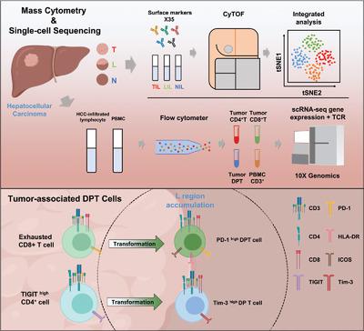
The spatial heterogeneity of immune microenvironment in hepatocellular carcinoma (HCC) remains elusive. Here, a single‐cell study involving 17 432 600 immune cells of 39 matched HCC (T), nontumor (N), and leading‐edge (L) specimens by mass cytometry is conducted. The tumor‐associated CD4/CD8 double‐positive T (DPT) cells are found enriched in L regions with synergetic expression of PD‐1/HLA‐DR/ICOS/CD45RO and exhibit a higher level of IFN‐γ , TNF‐α , and PD‐1 upon stimulation. The enrichment of DPT and PD‐1+DPT in L regions indicates favorable prognosis. These tumor‐associated DPT cells with similar phenotype are also verified in other tumors and HCC animal models. Single‐cell RNA‐seq further characterizes the molecular features of DPT cells and uncovers 11 clusters with different cytotoxicity, exhaustion, and activation scores. TCR‐based trajectory analysis reveals that tumor‐associated DPT clusters share separated ancestries with local CD4+ or CD8+SPT cells rather than CD3+PBMC cells. TCR clones with frequency above 10 are mainly found coexisting in DPT and CD8+SPT cells. Specifically, PD‐1highDPT cluster (TDPT_10) shares the same ancestry with exhausted CD8+SPT cluster (TCD8T_2) and shows higher expression similarity and closer pathological location to PD‐1+CD8+ than PD‐1+CD4+T cells. Together, this study systematically characterizes the unique distribution of PD‐1+DPTs in HCC and puts forward new insights for the function and origin of tumor‐associated DPT cells.
中文翻译:

通过单细胞细胞计数和转录组测序对肝细胞癌中 PD-1high CD4+CD8+ T 细胞的轨迹和功能进行分析。
肝细胞癌(HCC)免疫微环境的空间异质性仍然难以捉摸。在这里,通过质谱流式细胞仪进行了一项单细胞研究,涉及 39 个匹配的 HCC (T)、非肿瘤 (N) 和前沿 (L) 样本的 17 432 600 个免疫细胞。肿瘤相关 CD4/CD8 双阳性 T (DPT) 细胞在 L 区富集,协同表达 PD-1/HLA-DR/ICOS/CD45RO,并表现出较高水平的 IFN- γ 、TNF- α 、和PD-1刺激后。 L 区 DPT 和 PD-1 + DPT 富集表明预后良好。这些具有相似表型的肿瘤相关 DPT 细胞也在其他肿瘤和 HCC 动物模型中得到了验证。单细胞 RNA-seq 进一步表征了 DPT 细胞的分子特征,并揭示了 11 个具有不同细胞毒性、耗竭和激活评分的簇。基于 TCR 的轨迹分析表明,肿瘤相关 DPT 簇与局部 CD4 +或 CD8 + SPT 细胞而不是 CD3 + PBMC 细胞共享不同的祖先。频率在10以上的TCR克隆主要存在于DPT和CD8 + SPT细胞中。具体而言,PD-1高DPT簇(TDPT_10)与耗尽的CD8 + SPT簇(TCD8T_2)具有相同的祖先,并且与PD-1 + CD8 +相比,PD-1 + CD4 + T细胞表现出更高的表达相似性和更接近的病理位置。总之,这项研究系统地表征了 HCC 中 PD-1 + DPT 的独特分布,并对肿瘤相关 DPT 细胞的功能和起源提出了新的见解。
更新日期:2020-07-08
中文翻译:
通过单细胞细胞计数和转录组测序对肝细胞癌中 PD-1high CD4+CD8+ T 细胞的轨迹和功能进行分析。
肝细胞癌(HCC)免疫微环境的空间异质性仍然难以捉摸。在这里,通过质谱流式细胞仪进行了一项单细胞研究,涉及 39 个匹配的 HCC (T)、非肿瘤 (N) 和前沿 (L) 样本的 17 432 600 个免疫细胞。肿瘤相关 CD4/CD8 双阳性 T (DPT) 细胞在 L 区富集,协同表达 PD-1/HLA-DR/ICOS/CD45RO,并表现出较高水平的 IFN- γ 、TNF- α 、和PD-1刺激后。 L 区 DPT 和 PD-1 + DPT 富集表明预后良好。这些具有相似表型的肿瘤相关 DPT 细胞也在其他肿瘤和 HCC 动物模型中得到了验证。单细胞 RNA-seq 进一步表征了 DPT 细胞的分子特征,并揭示了 11 个具有不同细胞毒性、耗竭和激活评分的簇。基于 TCR 的轨迹分析表明,肿瘤相关 DPT 簇与局部 CD4 +或 CD8 + SPT 细胞而不是 CD3 + PBMC 细胞共享不同的祖先。频率在10以上的TCR克隆主要存在于DPT和CD8 + SPT细胞中。具体而言,PD-1高DPT簇(TDPT_10)与耗尽的CD8 + SPT簇(TCD8T_2)具有相同的祖先,并且与PD-1 + CD8 +相比,PD-1 + CD4 + T细胞表现出更高的表达相似性和更接近的病理位置。总之,这项研究系统地表征了 HCC 中 PD-1 + DPT 的独特分布,并对肿瘤相关 DPT 细胞的功能和起源提出了新的见解。


 京公网安备 11010802027423号