当前位置:
X-MOL 学术
›
J. Biophotonics
›
论文详情
Our official English website, www.x-mol.net, welcomes your
feedback! (Note: you will need to create a separate account there.)
Detection of oral squamous cell carcinoma with ex vivo fluorescence confocal microscopy: Sensitivity and specificity compared to histopathology.
Journal of Biophotonics ( IF 2.0 ) Pub Date : 2020-06-29 , DOI: 10.1002/jbio.202000100 Veronika Shavlokhova 1 , Christa Flechtenmacher 2 , Sameena Sandhu 1 , Maximilian Pilz 3 , Michael Vollmer 1 , Jürgen Hoffmann 1 , Michel Engel 1 , Christian Freudlsperger 1
Journal of Biophotonics ( IF 2.0 ) Pub Date : 2020-06-29 , DOI: 10.1002/jbio.202000100 Veronika Shavlokhova 1 , Christa Flechtenmacher 2 , Sameena Sandhu 1 , Maximilian Pilz 3 , Michael Vollmer 1 , Jürgen Hoffmann 1 , Michel Engel 1 , Christian Freudlsperger 1
Affiliation

|
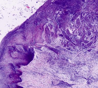
Real‐time microscopic imaging of freshly excised tissue enables a rapid bedside‐pathology. A possible application of interest is the detection of oral squamous cell carcinomas (OSCCs). The aim of this study was to analyze the sensitivity and specificity of ex vivo fluorescence confocal microscopy (FCM) for OSCCs and to compare confocal images visually and qualitatively with gold standard histopathology. Two hundred eighty ex vivo FCM images were prospectively collected and evaluated immediately after excision. Every confocal image was blindly assessed for the presence or absence of malignancy by two clinicians and one pathologist. The results were compared with conventional histopathology with hematoxylin and eosin staining. OSCCs were detected with a very high sensitivity of 0.991, specificity of 0.9527, positive predictive value of 0.9322 and negative predictive value of 0.9938. The results demonstrate the potential of ex vivo FCM in fresh tissue for rapid real‐time surgical pathology.
中文翻译:

用离体荧光共聚焦显微镜检测口腔鳞状细胞癌:与组织病理学相比,灵敏度和特异性更高。
新近切除的组织的实时显微成像可实现快速的床旁病理检查。感兴趣的可能应用是口腔鳞状细胞癌(OSCC)的检测。这项研究的目的是分析离体荧光共聚焦显微镜(FCM)对OSCC的敏感性和特异性,并在视觉和质量上与金标准组织病理学比较共聚焦图像。切除后立即收集并评估了280张离体FCM图像。由两名临床医生和一名病理医生盲目评估每个共聚焦图像是否存在恶性肿瘤。将结果与苏木精和曙红染色的常规组织病理学进行比较。检测到的OSCC的灵敏度非常高,为0.991,特异性为0.9527,阳性预测值为0。9322和0.9938的阴性预测值。结果表明,离体FCM在新鲜组织中具有快速实时手术病理学的潜力。
更新日期:2020-06-29

中文翻译:

用离体荧光共聚焦显微镜检测口腔鳞状细胞癌:与组织病理学相比,灵敏度和特异性更高。
新近切除的组织的实时显微成像可实现快速的床旁病理检查。感兴趣的可能应用是口腔鳞状细胞癌(OSCC)的检测。这项研究的目的是分析离体荧光共聚焦显微镜(FCM)对OSCC的敏感性和特异性,并在视觉和质量上与金标准组织病理学比较共聚焦图像。切除后立即收集并评估了280张离体FCM图像。由两名临床医生和一名病理医生盲目评估每个共聚焦图像是否存在恶性肿瘤。将结果与苏木精和曙红染色的常规组织病理学进行比较。检测到的OSCC的灵敏度非常高,为0.991,特异性为0.9527,阳性预测值为0。9322和0.9938的阴性预测值。结果表明,离体FCM在新鲜组织中具有快速实时手术病理学的潜力。


 京公网安备 11010802027423号