当前位置:
X-MOL 学术
›
J. Morphol.
›
论文详情
Our official English website, www.x-mol.net, welcomes your feedback! (Note: you will need to create a separate account there.)
Ultrastructural evidence for the origin of the subretinal pigment shield in the compound eye of Drosophila melanogaster
Journal of Morphology ( IF 1.5 ) Pub Date : 2020-05-12 , DOI: 10.1002/jmor.21143 Tobias Mohr 1, 2 , Stefan Fischer 2
Journal of Morphology ( IF 1.5 ) Pub Date : 2020-05-12 , DOI: 10.1002/jmor.21143 Tobias Mohr 1, 2 , Stefan Fischer 2
Affiliation
|
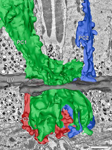
Little morphological information is available about subretinal pigment shields in insect compound eyes, especially ultrastructural information. The latter is however needed in order to detect possible smallest projections that emanate from pigment‐granule‐bearing cells and pass through the basal matrix (BM), but that are not visible in light micrographs. Previous work on the subretinal pigment shield in Drosophila melanogaster suggests that the pigment cell population located below the BM is closely associated with secondary and tertiary pigment cells. Whether these cells stay in connection throughout life with the subretinal regions via thin projections that pass through the fenestrae of the BM, or whether the subretinal parts later become separated during eye development remained so far unknown. Our investigation of the periphery of the BM by three‐dimensional reconstruction based on serial‐sectioning transmission electron microscopy has revealed that the secondary and tertiary pigment cells possess thin projections that pass through the fenestrae of the BM and thus connect the cellular regions above and below the BM in the adult compound eye. The subretinal pigment shield of D. melanogaster is therefore of retinal origin and is not composed of additional subretinal pigment cells. The maintained bond allows the active displacement of pigment granules below the BM during the process of dark and light adaptation of the compound eye.
中文翻译:

黑腹果蝇复眼视网膜下色素盾起源的超微结构证据
关于昆虫复眼视网膜下色素盾的形态学信息很少,尤其是超微结构信息。然而,需要后者来检测可能从带有色素颗粒的细胞发出并穿过基底基质 (BM) 的最小投影,但在光学显微照片中是不可见的。先前对黑腹果蝇视网膜下色素屏障的研究表明,位于 BM 下方的色素细胞群与二级和三级色素细胞密切相关。这些细胞是否在整个生命过程中通过穿过 BM 窗孔的薄突出物与视网膜下区域保持连接,或者视网膜下部分后来在眼睛发育过程中是否分离仍然未知。我们通过基于连续切片透射电子显微镜的三维重建对 BM 周边的研究表明,二级和三级色素细胞具有穿过 BM 窗孔的薄突起,从而连接上方和下方的细胞区域成人复眼中的 BM。因此,黑腹果蝇的视网膜下色素盾是视网膜起源的,不是由额外的视网膜下色素细胞组成。在复眼的明暗适应过程中,保持的键允许BM下方的色素颗粒主动位移。
更新日期:2020-05-12
中文翻译:
黑腹果蝇复眼视网膜下色素盾起源的超微结构证据
关于昆虫复眼视网膜下色素盾的形态学信息很少,尤其是超微结构信息。然而,需要后者来检测可能从带有色素颗粒的细胞发出并穿过基底基质 (BM) 的最小投影,但在光学显微照片中是不可见的。先前对黑腹果蝇视网膜下色素屏障的研究表明,位于 BM 下方的色素细胞群与二级和三级色素细胞密切相关。这些细胞是否在整个生命过程中通过穿过 BM 窗孔的薄突出物与视网膜下区域保持连接,或者视网膜下部分后来在眼睛发育过程中是否分离仍然未知。我们通过基于连续切片透射电子显微镜的三维重建对 BM 周边的研究表明,二级和三级色素细胞具有穿过 BM 窗孔的薄突起,从而连接上方和下方的细胞区域成人复眼中的 BM。因此,黑腹果蝇的视网膜下色素盾是视网膜起源的,不是由额外的视网膜下色素细胞组成。在复眼的明暗适应过程中,保持的键允许BM下方的色素颗粒主动位移。


 京公网安备 11010802027423号