当前位置:
X-MOL 学术
›
J. Biophotonics
›
论文详情
Our official English website, www.x-mol.net, welcomes your
feedback! (Note: you will need to create a separate account there.)
Vascular changes precede tomographic changes in diabetic eyes without retinopathy and improve artificial intelligence diagnostics.
Journal of Biophotonics ( IF 2.0 ) Pub Date : 2020-06-18 , DOI: 10.1002/jbio.202000107 Nivedhitha Govindaswamy 1 , Dhanashree Ratra 2 , Daleena Dalan 2 , Subashchandra Doralli 3 , Anirudha A Tirumalai 3 , Rajesh Nagarajan 2 , Thirumalesh Mochi 3 , Naren Shetty 4 , Abhijit Sinha Roy 1
Journal of Biophotonics ( IF 2.0 ) Pub Date : 2020-06-18 , DOI: 10.1002/jbio.202000107 Nivedhitha Govindaswamy 1 , Dhanashree Ratra 2 , Daleena Dalan 2 , Subashchandra Doralli 3 , Anirudha A Tirumalai 3 , Rajesh Nagarajan 2 , Thirumalesh Mochi 3 , Naren Shetty 4 , Abhijit Sinha Roy 1
Affiliation
|
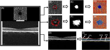
The purpose of this study was to evaluate early vascular and tomographic changes in the retina of diabetic patients using artificial intelligence (AI). The study included 74 age‐matched normal eyes, 171 diabetic eyes without retinopathy (DWR) eyes and 69 mild non‐proliferative diabetic retinopathy (NPDR) eyes. All patients underwent optical coherence tomography angiography (OCTA) imaging. Tomographic features (thickness and volume) were derived from the OCTA B‐scans. These features were used in AI models. Both OCT and OCTA features showed significant differences between the groups (P < .05). However, the OCTA features indicated early retinal changes in DWR eyes better than OCT (P < .05). In the AI model using both OCT and OCTA features simultaneously, the best area under the curve of 0.91 ± 0.02 was obtained (P < .05). Thus, the combined use of AI, OCT and OCTA significantly improved the early diagnosis of diabetic changes in the retina.
中文翻译:

在没有视网膜病变的情况下,糖尿病眼中的血管变化先于断层扫描,并改善了人工智能诊断。
这项研究的目的是使用人工智能(AI)评估糖尿病患者视网膜的早期血管和断层扫描变化。该研究包括74例年龄相匹配的正常眼,171例无视网膜病变(DWR)的糖尿病眼和69例轻度非增生性糖尿病视网膜病变(NPDR)眼。所有患者均接受了光学相干断层扫描血管造影(OCTA)成像。断层扫描特征(厚度和体积)来自OCTA B扫描。这些功能已在AI模型中使用。OCT和OCTA的功能在两组之间均显示出显着差异(P <.05)。然而,OCTA的特征表明DWR眼的早期视网膜变化优于OCT(P<.05)。在同时使用OCT和OCTA功能的AI模型中,曲线下的最佳面积为0.91±0.02(P <.05)。因此,AI,OCT和OCTA的联合使用显着改善了视网膜中糖尿病变化的早期诊断。
更新日期:2020-06-18
中文翻译:
在没有视网膜病变的情况下,糖尿病眼中的血管变化先于断层扫描,并改善了人工智能诊断。
这项研究的目的是使用人工智能(AI)评估糖尿病患者视网膜的早期血管和断层扫描变化。该研究包括74例年龄相匹配的正常眼,171例无视网膜病变(DWR)的糖尿病眼和69例轻度非增生性糖尿病视网膜病变(NPDR)眼。所有患者均接受了光学相干断层扫描血管造影(OCTA)成像。断层扫描特征(厚度和体积)来自OCTA B扫描。这些功能已在AI模型中使用。OCT和OCTA的功能在两组之间均显示出显着差异(P <.05)。然而,OCTA的特征表明DWR眼的早期视网膜变化优于OCT(P<.05)。在同时使用OCT和OCTA功能的AI模型中,曲线下的最佳面积为0.91±0.02(P <.05)。因此,AI,OCT和OCTA的联合使用显着改善了视网膜中糖尿病变化的早期诊断。


 京公网安备 11010802027423号