当前位置:
X-MOL 学术
›
J. Biophotonics
›
论文详情
Our official English website, www.x-mol.net, welcomes your
feedback! (Note: you will need to create a separate account there.)
Colon cancer detection by using Poincaré sphere and 2D polarimetric mapping of ex vivo colon samples.
Journal of Biophotonics ( IF 2.0 ) Pub Date : 2020-05-28 , DOI: 10.1002/jbio.202000082 Deyan Ivanov 1 , Viktor Dremin 2, 3 , Alexander Bykov 3 , Ekaterina Borisova 1, 4 , Tsanislava Genova 1 , Alexey Popov 5 , Razvigor Ossikovski 6 , Tatiana Novikova 6 , Igor Meglinski 3, 7, 8, 9, 10
Journal of Biophotonics ( IF 2.0 ) Pub Date : 2020-05-28 , DOI: 10.1002/jbio.202000082 Deyan Ivanov 1 , Viktor Dremin 2, 3 , Alexander Bykov 3 , Ekaterina Borisova 1, 4 , Tsanislava Genova 1 , Alexey Popov 5 , Razvigor Ossikovski 6 , Tatiana Novikova 6 , Igor Meglinski 3, 7, 8, 9, 10
Affiliation
|
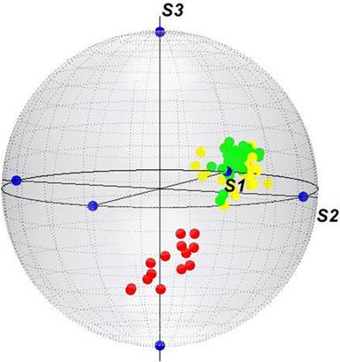
This work is dedicated to the diagnosis and grading of colon cancer by a combined use of Poincaré sphere and 2D Stokes vector polarimetry mapping approaches. The major challenge consists in exploring the applicability of polarized light for noninvasive screening of the histological abnormalities within the samples of biological tissues. Experimental studies were conducted in ex vivo colon sample, excised after surgical procedure for colon tumor removal of G2‐adenocarcinoma lesion. Polarimetric measurements in linear and circular regime were carried via personally developed polarimetric, optical set‐up, using supercontinuous fiber laser with irradiation fixed at 635 nm. We apply the Poincaré sphere and two‐dimensional Stokes vector scanning approach for screening the corresponding tissue samples. A comparison between linear and circular polarization states is made both for quantitative and qualitative evaluations. It is shown that circular polarization has better diagnostic capabilities than linear polarization, with higher dynamic ranges of the polarimetric parameters and better values of the diagnostic quantities. In addition to the standard polarimetry parameters, utilized as essential diagnostic markers, we apply statistical analysis to obtain more detailed information in frame of the applied diagnostic approach.
中文翻译:

通过使用庞加莱球和离体结肠样本的二维极化图检测结肠癌。
这项工作致力于通过结合使用庞加莱球和2D Stokes矢量极化法映射方法,对结肠癌进行诊断和分级。主要挑战在于探索偏振光在生物组织样品内组织学异常的无创筛查中的适用性。在离体结肠样本中进行了实验研究,该样本是在外科手术后切除以切除G2腺癌病变的结肠肿瘤。使用超连续光纤激光器,通过固定在635 nm的辐射,通过个人开发的偏振,光学设置进行了线性和圆形状态下的偏振测量。我们应用庞加莱球和二维斯托克斯矢量扫描方法来筛选相应的组织样本。进行线性和圆极化状态之间的比较,以进行定量和定性评估。结果表明,圆极化比线性极化具有更好的诊断能力,极化参数的动态范围更高,诊断量值更高。除了标准的旋光度参数(用作必要的诊断标记)外,我们还应用统计分析以获取所应用诊断方法框架内的更多详细信息。
更新日期:2020-05-28
中文翻译:
通过使用庞加莱球和离体结肠样本的二维极化图检测结肠癌。
这项工作致力于通过结合使用庞加莱球和2D Stokes矢量极化法映射方法,对结肠癌进行诊断和分级。主要挑战在于探索偏振光在生物组织样品内组织学异常的无创筛查中的适用性。在离体结肠样本中进行了实验研究,该样本是在外科手术后切除以切除G2腺癌病变的结肠肿瘤。使用超连续光纤激光器,通过固定在635 nm的辐射,通过个人开发的偏振,光学设置进行了线性和圆形状态下的偏振测量。我们应用庞加莱球和二维斯托克斯矢量扫描方法来筛选相应的组织样本。进行线性和圆极化状态之间的比较,以进行定量和定性评估。结果表明,圆极化比线性极化具有更好的诊断能力,极化参数的动态范围更高,诊断量值更高。除了标准的旋光度参数(用作必要的诊断标记)外,我们还应用统计分析以获取所应用诊断方法框架内的更多详细信息。


 京公网安备 11010802027423号