Journal of Neuroscience Methods ( IF 2.7 ) Pub Date : 2020-05-11 , DOI: 10.1016/j.jneumeth.2020.108765 Philip Anner 1 , Johannes Passecker 2 , Thomas Klausberger 2 , Georg Dorffner 3
|
Background
Cognitive neuroscientists aim to understand behavior often based on the underlying activity of individual neurons. Recently developed miniaturized epifluorescence microscopes allow recording of cellular calcium transients, resembling neuronal activity, of individual neurons even in deep brain areas in freely behaving animals. At the same time, molecular markers allow the characterization of diverse neuronal subtypes by post hoc immunohistochemical labeling. Combining both methods would allow researchers to increase insights into how individual neuronal activity and entities contribute to behavior.
New method
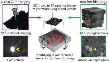
Here, we present a novel method for identifying the same neurons, recorded with calcium imaging using a miniaturized epifluorescence microscope, post hoc in fixed histological sections. This allows immunohistochemical investigations to detect the molecular signature of in vivo recorded neurons. Our method utilizes the structure of blood vessels for aligning in vivo acquired 2D images with a reconstructed 3D histological model.
Results
We automatically matched, 60 % of all in vivo recorded cells post hoc in histology. Across all animals, we successfully matched 43 % to 89 % of the recorded neurons. We provide a measure for the confidence of matched cells and validated our method by multiple simulation studies.
Comparison with existing methods
To our knowledge, we present the first method for matching cells, recorded with a miniaturized epifluorescence microscope in freely moving animals, post hoc in histological sections.
Conclusions
Our method allows a comprehensive analysis of how cortical circuits relate to freely moving animal behavior by combining functional activity of individual neurons with their underlying histological profiles.
中文翻译:
自动事后组织学鉴定自由活动大鼠神经元的Ca2 +成像。
背景
认知神经科学家的目标是经常基于单个神经元的潜在活动来理解行为。最近开发的微型落射荧光显微镜可以记录单个神经元的细胞钙瞬变,类似于神经元活动,即使在行为自由的动物的大脑深处也是如此。同时,分子标记可以通过事后免疫组织化学标记表征多种神经元亚型。两种方法的结合将使研究人员能够更深入地了解个体神经元活动和实体如何促进行为。
新方法
在这里,我们提出了一种用于识别相同神经元的新方法,该方法是使用固定的组织学切片在事后用小型表荧光显微镜的钙成像记录的。这使得免疫组织化学研究可以检测体内记录的神经元的分子标记。我们的方法利用血管结构将体内获得的2D图像与重建的3D组织学模型对齐。
结果
我们自动匹配,所有的60%,在体内记录细胞事后组织学。在所有动物中,我们成功地匹配了43%至89%的记录神经元。我们提供了对匹配单元格置信度的度量,并通过多次仿真研究验证了我们的方法。
与现有方法的比较
据我们所知,我们目前用于匹配单元的第一种方法,在自由活动动物的微型落射荧光显微镜记录,事后组织切片。
结论
我们的方法可以通过将单个神经元的功能活动与其潜在的组织学特征相结合,对皮质回路与自由移动的动物行为之间的关系进行全面分析。


 京公网安备 11010802027423号