当前位置:
X-MOL 学术
›
J. Synchrotron Radiat.
›
论文详情
Our official English website, www.x-mol.net, welcomes your
feedback! (Note: you will need to create a separate account there.)
Synchrotron multimodal imaging in a whole cell reveals lipid droplet core organization.
Journal of Synchrotron Radiation ( IF 2.4 ) Pub Date : 2020-04-23 , DOI: 10.1107/s1600577520003847 Frédéric Jamme 1 , Bertrand Cinquin 1 , Yann Gohon 2 , Eva Pereiro 3 , Matthieu Réfrégiers 1 , Marine Froissard 2
Journal of Synchrotron Radiation ( IF 2.4 ) Pub Date : 2020-04-23 , DOI: 10.1107/s1600577520003847 Frédéric Jamme 1 , Bertrand Cinquin 1 , Yann Gohon 2 , Eva Pereiro 3 , Matthieu Réfrégiers 1 , Marine Froissard 2
Affiliation
|
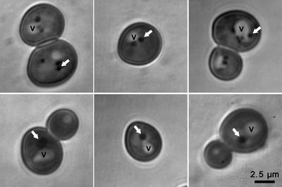
A lipid droplet (LD) core of a cell consists mainly of neutral lipids, triacylglycerols and/or steryl esters (SEs). The structuration of these lipids inside the core is still under debate. Lipid segregation inside LDs has been observed but is sometimes suggested to be an artefact of LD isolation and chemical fixation. LD imaging in their native state and in unaltered cellular environments appears essential to overcome these possible technical pitfalls. Here, imaging techniques for ultrastructural study of native LDs in cellulo are provided and it is shown that LDs are organized structures. Cryo soft X-ray tomography and deep-ultraviolet (DUV) transmittance imaging are showing a partitioning of SEs at the periphery of the LD core. Furthermore, DUV transmittance and tryptophan/tyrosine auto-fluorescence imaging on living cells are combined to obtain complementary information on cell chemical contents. This multimodal approach paves the way for a new label-free organelle imaging technique in living cells.
中文翻译:

全细胞同步加速器多模态成像揭示了脂滴核心组织。
细胞的脂滴(LD)核心主要由中性脂质、三酰甘油和/或甾醇酯(SE)组成。核心内这些脂质的结构仍然存在争议。已观察到 LD 内的脂质分离,但有时认为这是 LD 分离和化学固定的人为因素。天然状态和未改变的细胞环境中的 LD 成像对于克服这些可能的技术缺陷至关重要。在这里,提供了用于细胞中天然 LD 的超微结构研究的成像技术,并且表明 LD 是有组织的结构。低温软 X 射线断层扫描和深紫外 (DUV) 透射率成像显示 LD 核心外围有 SE 分区。此外,将活细胞的深紫外透射率和色氨酸/酪氨酸自发荧光成像相结合,以获得细胞化学含量的补充信息。这种多模式方法为活细胞中新的无标记细胞器成像技术铺平了道路。
更新日期:2020-04-23
中文翻译:
全细胞同步加速器多模态成像揭示了脂滴核心组织。
细胞的脂滴(LD)核心主要由中性脂质、三酰甘油和/或甾醇酯(SE)组成。核心内这些脂质的结构仍然存在争议。已观察到 LD 内的脂质分离,但有时认为这是 LD 分离和化学固定的人为因素。天然状态和未改变的细胞环境中的 LD 成像对于克服这些可能的技术缺陷至关重要。在这里,提供了用于细胞中天然 LD 的超微结构研究的成像技术,并且表明 LD 是有组织的结构。低温软 X 射线断层扫描和深紫外 (DUV) 透射率成像显示 LD 核心外围有 SE 分区。此外,将活细胞的深紫外透射率和色氨酸/酪氨酸自发荧光成像相结合,以获得细胞化学含量的补充信息。这种多模式方法为活细胞中新的无标记细胞器成像技术铺平了道路。


 京公网安备 11010802027423号