Methods ( IF 4.2 ) Pub Date : 2020-05-07 , DOI: 10.1016/j.ymeth.2020.05.004 Mark S Schröder 1 , Marie-Lena I E Harwardt 1 , Johanna V Rahm 1 , Yunqing Li 1 , Petra Freund 1 , Marina S Dietz 1 , Mike Heilemann 1
|
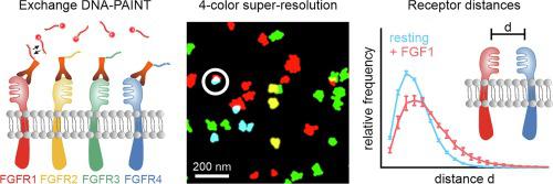
Fibroblast growth factor receptors (FGFRs) are a subfamily of receptor tyrosine kinases and central players in health and disease. Following ligand binding and the formation of homo- and heteromeric complexes, FGFRs initiate a cellular response. Challenges in studying FGFR activation are inner-subfamily interactions and a complex heterogeneity of these in the cell membrane, which demand for observation techniques that can resolve individual protein complexes and that are compatible with endogenous protein levels. Here, we established an imaging and analysis pipeline for multiplexed single-molecule localization microscopy (SMLM) of the FGFR network at the plasma membrane. Using DNA-labeled primary antibodies, we visualize all four FGFRs in the same cell with near-molecular spatial resolution. From the super-resolution imaging data, we extract information on FGFR density, spatial distribution, and inner-subfamily colocalization. Our approach is straightforward and easily adaptable to other multiplexed SMLM data of membrane proteins.
中文翻译:
用 DNA 辅助单分子超分辨率显微镜对质膜上的成纤维细胞生长因子受体网络进行成像
成纤维细胞生长因子受体 (FGFR) 是受体酪氨酸激酶的一个亚家族,是健康和疾病的核心参与者。在配体结合并形成同型和异型复合物后,FGFR 会启动细胞反应。研究 FGFR 激活的挑战是内亚家族相互作用和细胞膜中这些相互作用的复杂异质性,这需要能够解析单个蛋白质复合物并与内源性蛋白质水平兼容的观察技术。在这里,我们为质膜处的 FGFR 网络的多路复用单分子定位显微镜 (SMLM) 建立了成像和分析管道。使用 DNA 标记的一抗,我们以近分子的空间分辨率可视化同一细胞中的所有四个 FGFR。从超分辨率成像数据来看,我们提取有关 FGFR 密度、空间分布和内部亚科共定位的信息。我们的方法很简单,很容易适应膜蛋白的其他多路复用 SMLM 数据。


 京公网安备 11010802027423号