当前位置:
X-MOL 学术
›
Ultramicroscopy
›
论文详情
Our official English website, www.x-mol.net, welcomes your
feedback! (Note: you will need to create a separate account there.)
Integrated super resolution fluorescence microscopy and transmission electron microscopy
Ultramicroscopy ( IF 2.1 ) Pub Date : 2020-08-01 , DOI: 10.1016/j.ultramic.2020.113007 Sajjad Mohammadian 1 , Alexandra V Agronskaia 1 , Gerhard A Blab 1 , Elly G van Donselaar 2 , Cecilia de Heus 2 , Nalan Liv 2 , Judith Klumperman 2 , Hans C Gerritsen 1
Ultramicroscopy ( IF 2.1 ) Pub Date : 2020-08-01 , DOI: 10.1016/j.ultramic.2020.113007 Sajjad Mohammadian 1 , Alexandra V Agronskaia 1 , Gerhard A Blab 1 , Elly G van Donselaar 2 , Cecilia de Heus 2 , Nalan Liv 2 , Judith Klumperman 2 , Hans C Gerritsen 1
Affiliation
|
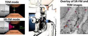
In correlative light and electron microscopy (CLEM), the capabilities of fluorescence microscopy (FM) and electron microscopy (EM) are united. FM combines a large field of view with high sensitivity for detecting fluorescence, which makes it an excellent tool for identifying regions of interest. EM has a much smaller field of view but offers superb resolution that allows studying cellular ultrastructure. In CLEM, the potentials of both techniques are combined but a limiting factor is the large difference in resolution between the two imaging modalities. Adding super resolution FM to CLEM reduces the resolution gap between FM and EM; it offers the possibility of identifying multiple targets within the diffraction limit and can increase correlation accuracy. CLEM is usually carried out in two separate setups, which requires transfer of the sample. This may result in distortion and damage of the specimen, which can complicate finding back regions of interest. By integrating the two imaging modalities, such problems can be avoided. Here, an integrated super resolution correlative microscopy approach is presented based on a wide-field super resolution FM integrated in a Transmission Electron Microscope (TEM). Switching imaging modalities is accomplished by rotation of the TEM sample holder. First imaging experiments are presented on sections of Lowicryl embedded Human Umbilical Vein Endothelial Cells labeled for Caveolin both with Protein A-Gold, and Alexa Fluor®647. TEM and FM images were overlaid using fiducial markers visible in both imaging modalities with an overlay accuracy of 28 ± 11 nm. This is close to the optical resolution of ~50 nm.
中文翻译:

集成超分辨率荧光显微镜和透射电子显微镜
在相关光电子显微镜 (CLEM) 中,荧光显微镜 (FM) 和电子显微镜 (EM) 的功能结合在一起。FM 结合了大视野和检测荧光的高灵敏度,这使其成为识别感兴趣区域的绝佳工具。EM 的视野要小得多,但提供了极好的分辨率,可以研究细胞超微结构。在 CLEM 中,结合了两种技术的潜力,但一个限制因素是两种成像方式之间的分辨率差异很大。CLEM加入超分辨率FM,缩小FM和EM的分辨率差距;它提供了在衍射极限内识别多个目标的可能性,并可以提高相关精度。CLEM 通常在两个独立的设置中进行,这需要转移样品。这可能会导致标本的变形和损坏,这会使寻找感兴趣的区域变得复杂。通过集成两种成像方式,可以避免此类问题。在这里,基于集成在透射电子显微镜 (TEM) 中的宽场超分辨率 FM,提出了一种集成的超分辨率相关显微镜方法。切换成像模式是通过旋转 TEM 样品架来完成的。首次成像实验展示在 Lowicryl 嵌入的人脐静脉内皮细胞切片上,用蛋白 A-Gold 和 Alexa Fluor®647 标记了小窝蛋白。TEM 和 FM 图像使用在两种成像模式中可见的基准标记重叠,重叠精度为 28 ± 11 nm。这接近于~50 nm 的光学分辨率。
更新日期:2020-08-01
中文翻译:
集成超分辨率荧光显微镜和透射电子显微镜
在相关光电子显微镜 (CLEM) 中,荧光显微镜 (FM) 和电子显微镜 (EM) 的功能结合在一起。FM 结合了大视野和检测荧光的高灵敏度,这使其成为识别感兴趣区域的绝佳工具。EM 的视野要小得多,但提供了极好的分辨率,可以研究细胞超微结构。在 CLEM 中,结合了两种技术的潜力,但一个限制因素是两种成像方式之间的分辨率差异很大。CLEM加入超分辨率FM,缩小FM和EM的分辨率差距;它提供了在衍射极限内识别多个目标的可能性,并可以提高相关精度。CLEM 通常在两个独立的设置中进行,这需要转移样品。这可能会导致标本的变形和损坏,这会使寻找感兴趣的区域变得复杂。通过集成两种成像方式,可以避免此类问题。在这里,基于集成在透射电子显微镜 (TEM) 中的宽场超分辨率 FM,提出了一种集成的超分辨率相关显微镜方法。切换成像模式是通过旋转 TEM 样品架来完成的。首次成像实验展示在 Lowicryl 嵌入的人脐静脉内皮细胞切片上,用蛋白 A-Gold 和 Alexa Fluor®647 标记了小窝蛋白。TEM 和 FM 图像使用在两种成像模式中可见的基准标记重叠,重叠精度为 28 ± 11 nm。这接近于~50 nm 的光学分辨率。


 京公网安备 11010802027423号