IRBM ( IF 5.6 ) Pub Date : 2020-02-29 , DOI: 10.1016/j.irbm.2020.02.002 M. Mohtasebi , M. Bayat , S. Ghadimi , H. Abrishami Moghaddam , F. Wallois

|
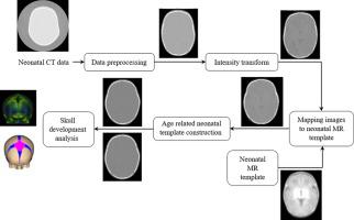
Neonatal skull anatomy and its evolution have received less attention with respect to the brain anatomy in neuroscience and neuroanatomy studies. Meanwhile, their influence on normal brain development and their impact on the results of functional brain studies have been demonstrated by several researches. Such disesteem is due to the weak appearance of the cranial bones, fontanels and sutures in images acquired by MRI which presents actually the only available aperture for observing the neonatal head volume in details. This paper presents an unprecedented retrospective CT-based study on modeling the neonatal skull and its development during the first weeks of life in a standard space defined by the available neonatal MRI model. We create two neonatal head atlases for the age ranges of 39-40 and 41-42 week's gestational age using symmetric group-wise normalization method. The created atlases allow direct observation of ossification patterns and precise three-dimensional measurement of anatomical features from neonatal skull during development. Development of the neonatal skull has been examined here using nineteen CT scans of neonates with two-week gestational age ranges of 39 to 40 and 41 to 42. Deformation-based morphometry method is applied with the use of Jacobian determinant maps to identify growth patterns and observe ossification during specified time interval. Precise three-dimensional measurements of anterior fontanel size, scalp eliminated head circumference and the area corresponding to the fontanel-sutures were performed by extracting fontanels and sutures.
中文翻译:

使用计算机断层扫描图像对新生儿头骨发育进行建模
在神经科学和神经解剖学研究中,关于脑部解剖学,新生儿颅骨解剖学及其发展受到的关注较少。同时,一些研究证明了它们对正常大脑发育的影响以及对功能性大脑研究结果的影响。这种厌恶是由于MRI采集的图像中颅骨,font石和缝合线的外观较弱,而MRI实际上是唯一可用来详细观察新生儿头颅容积的孔径。本文提出了一项基于CT的史无前例的回顾性研究,该研究在可用的新生儿MRI模型定义的标准空间中,对生命的头几周内新生儿头骨及其发育进行建模。我们创建了两个39-40周和41-42周龄的新生儿头部图集 使用对称分组归一化方法的胎龄。创建的地图集可直接观察骨化模式,并在发育过程中精确地从新生儿颅骨解剖特征进行三维测量。新生儿颅骨的发育已在这里进行了十九周两周胎龄为39至40岁和41至42岁的新生儿的CT扫描。基于变形的形态学方法与雅可比行列式图一起使用,以确定生长方式和在指定的时间间隔内观察骨化。通过提取font和缝线进行前font大小,头皮消除头围和与corresponding缝相对应的面积的精确三维测量。创建的地图集可直接观察骨化模式,并在发育过程中精确地从新生儿颅骨解剖特征进行三维测量。新生儿颅骨的发育已在这里进行了19周CT扫描,两周胎龄为39至40岁和41至42岁的新生儿进行了检查。基于变形的形态计量学方法与雅可比行列式图一起用于识别生长模式和在指定的时间间隔内观察骨化。通过提取font和缝线进行前font大小,头皮消除头围和与corresponding缝相对应的面积的精确三维测量。创建的地图集可直接观察骨化模式,并在发育过程中精确地从新生儿颅骨解剖特征进行三维测量。新生儿颅骨的发育已在这里进行了十九周两周胎龄为39至40岁和41至42岁的新生儿的CT扫描。基于变形的形态学方法与雅可比行列式图一起使用,以确定生长方式和在指定的时间间隔内观察骨化。通过提取font和缝线进行前font大小,头皮消除头围和与corresponding缝相对应的面积的精确三维测量。新生儿颅骨的发育已在这里进行了十九周两周胎龄为39至40岁和41至42岁的新生儿的CT扫描。基于变形的形态学方法与雅可比行列式图一起使用,以确定生长方式和在指定的时间间隔内观察骨化。通过提取font和缝线进行前font大小,头皮消除头围和与corresponding缝相对应的面积的精确三维测量。新生儿颅骨的发育已在这里进行了十九周两周胎龄为39至40岁和41至42岁的新生儿的CT扫描。基于变形的形态学方法与雅可比行列式图一起使用,以确定生长方式和在指定的时间间隔内观察骨化。通过提取font和缝线进行前font大小,头皮消除头围和与corresponding缝相对应的面积的精确三维测量。


 京公网安备 11010802027423号