当前位置:
X-MOL 学术
›
Bioconjugate Chem.
›
论文详情
Our official English website, www.x-mol.net, welcomes your
feedback! (Note: you will need to create a separate account there.)
Supramolecular Encapsulation of Small-Ultrared Fluorescent Proteins in Virus-Like Nanoparticles for Noninvasive In Vivo Imaging Agents.
Bioconjugate Chemistry ( IF 4.0 ) Pub Date : 2020-05-07 , DOI: 10.1021/acs.bioconjchem.0c00190 Fabian C. Herbert , Olivia R. Brohlin , Tyler Galbraith , Candace Benjamin , Cesar A. Reyes , Michael A. Luzuriaga , Arezoo Shahrivarkevishahi , Jeremiah J. Gassensmith
Bioconjugate Chemistry ( IF 4.0 ) Pub Date : 2020-05-07 , DOI: 10.1021/acs.bioconjchem.0c00190 Fabian C. Herbert , Olivia R. Brohlin , Tyler Galbraith , Candace Benjamin , Cesar A. Reyes , Michael A. Luzuriaga , Arezoo Shahrivarkevishahi , Jeremiah J. Gassensmith
|
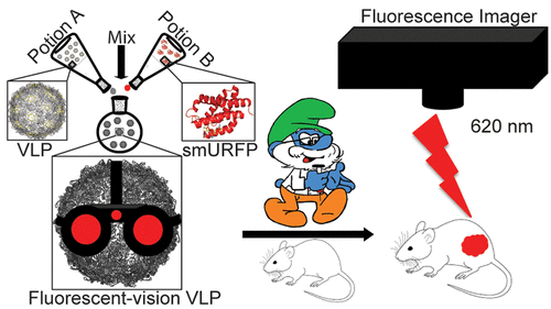
Icosahedral virus-like particles (VLPs) derived from bacteriophages Qβ and PP7 encapsulating small-ultrared fluorescent protein (smURFP) were produced using a versatile supramolecular capsid disassemble-reassemble approach. The generated fluorescent VLPs display identical structural properties to their nonfluorescent analogs. Encapsulated smURFP shows indistinguishable photochemical properties to its unencapsulated counterpart, exhibits outstanding stability toward pH, and produces bright in vitro images following phagocytosis by macrophages. In vivo imaging allows the biodistribution to be imaged at different time points. Ex vivo imaging of intravenously administered encapsulated smURFP reveals a localization in the liver and kidneys after 2 h blood circulation and substantial elimination after 16 h of imaging, highlighting the potential application of these constructs as noninvasive in vivo imaging agents.
中文翻译:

将小紫外荧光蛋白超分子封装在病毒样纳米颗粒中,用于无创体内成像剂。
使用通用的超分子衣壳拆装重组方法生产源自噬菌体 Qβ 和 PP7 的二十面体病毒样颗粒 (VLP),其封装小紫外荧光蛋白 (smURFP)。生成的荧光 VLP 显示出与其非荧光类似物相同的结构特性。封装的 smURFP 与其未封装的对应物表现出无法区分的光化学特性,对 pH 值表现出出色的稳定性,并在被巨噬细胞吞噬后产生明亮的体外图像。体内成像允许在不同时间点对生物分布进行成像。静脉注射封装的 smURFP 的离体成像揭示了 2 小时血液循环后在肝脏和肾脏中的定位,并在成像 16 小时后基本消除,突出了这些结构作为非侵入性体内成像剂的潜在应用。
更新日期:2020-04-28
中文翻译:
将小紫外荧光蛋白超分子封装在病毒样纳米颗粒中,用于无创体内成像剂。
使用通用的超分子衣壳拆装重组方法生产源自噬菌体 Qβ 和 PP7 的二十面体病毒样颗粒 (VLP),其封装小紫外荧光蛋白 (smURFP)。生成的荧光 VLP 显示出与其非荧光类似物相同的结构特性。封装的 smURFP 与其未封装的对应物表现出无法区分的光化学特性,对 pH 值表现出出色的稳定性,并在被巨噬细胞吞噬后产生明亮的体外图像。体内成像允许在不同时间点对生物分布进行成像。静脉注射封装的 smURFP 的离体成像揭示了 2 小时血液循环后在肝脏和肾脏中的定位,并在成像 16 小时后基本消除,突出了这些结构作为非侵入性体内成像剂的潜在应用。


 京公网安备 11010802027423号