当前位置:
X-MOL 学术
›
Cell Biol. Int.
›
论文详情
Our official English website, www.x-mol.net, welcomes your
feedback! (Note: you will need to create a separate account there.)
A comparative study of hBM-MSCs' differentiation toward osteogenic lineage in the presence of progesterone and estrogen hormones separately and concurrently in vitro.
Cell Biology International ( IF 3.3 ) Pub Date : 2020-04-27 , DOI: 10.1002/cbin.11364 Maryam Soltanyzadeh 1 , Marzieh Ghollasi 1 , Raheleh Halabian 2 , Mehdi Shams 3
Cell Biology International ( IF 3.3 ) Pub Date : 2020-04-27 , DOI: 10.1002/cbin.11364 Maryam Soltanyzadeh 1 , Marzieh Ghollasi 1 , Raheleh Halabian 2 , Mehdi Shams 3
Affiliation
|
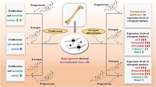
Promising cell sources for tissue engineering comprise bone marrow derived‐mesenchymal stem cells (BM‐MSCs) that have multiple differentiation potentials. Also, sex hormones act as important elements in bone development and maintenance, and the roles of two female sex steroid hormones known as estrogen (17‐β estradiol) and progesterone in osteogenic differentiation of human BM‐MSCs (hBM‐MSCs) are studied. For this purpose, hBM‐MSCs were treated with a 1 × 10⁻⁶ M concentration of 17‐β estradiol and progesterone separately and simultaneously while the optimum concentrations were obtained by 3‐(4,5‐dimethylthiazol‐2‐yl)‐2,5‐diphenyltetrazolium bromide (MTT) assay. Osteogenic differentiation tests including measurement of alkaline phosphatase (ALP) enzyme activity, the content of total mineral calcium, mineralized matrix staining by Alizarin Red and Von Kossa solutions, real‐time reverse transcription polymerase chain reaction (RT‐PCR), and immunofluorescence staining were carried out on Days 7 and 14 of differentiation. To exhibit the morphology of the cells, the BM‐MSCs were stained with acridine orange (AO) solution. In this study, the results of ALP activity assay, calcium content and real‐time RT‐PCR assay and also all tests of differentiation staining have shown that 17‐β estradiol has been recognized as an enhancing factor of osteogenic differentiation. Furthermore, MTT assay and AO staining revealed progesterone as a factor that seriously improved the proliferation of hBM‐MSCs. Generally, the 17‐β estradiol individually or in the presence of progesterone has more effects on BM‐MSCs' osteogenic differentiation compared to progesterone alone. In this study, it is indicated that the effect of the 17‐β estradiol and progesterone concurrently was the same as individual 17‐β estradiol on the differentiation of hBM‐MSCs.
中文翻译:

在体外和同时存在孕激素和雌激素的情况下,hBM-MSC向成骨谱系分化的比较研究。
用于组织工程的有希望的细胞来源包括具有多种分化潜能的骨髓衍生间充质干细胞(BM-MSC)。此外,性激素是骨骼发育和维持的重要元素,并且研究了两种雌激素类固醇激素(雌激素(17-β雌二醇)和孕酮)在人BM-MSC(hBM-MSC)的成骨分化中的作用。为此,将hBM-MSC用1×10 -6处理 分别和同时测定17-β雌二醇和孕酮的M浓度,而最佳浓度则通过3-(4,5-二甲基噻唑-2-基)-2,5-二苯基四唑溴化物(MTT)测定获得。成骨分化测试包括碱性磷酸酶(ALP)酶活性测量,总矿物质钙含量,茜素红和Von Kossa溶液对矿化基质染色,实时逆转录聚合酶链反应(RT-PCR)和免疫荧光染色在分化的第7和14天进行。为了展示细胞的形态,用‐啶橙(AO)溶液对BM-MSC进行染色。在这项研究中,ALP活性测定的结果 钙含量和实时RT-PCR分析以及所有分化染色测试均表明17-β雌二醇已被认为是成骨分化的增强因子。此外,MTT分析和AO染色显示孕酮是严重改善hBM-MSCs增殖的因素。通常,与单独使用孕激素相比,单独使用17-β雌二醇或在孕酮存在下对雌激素的影响更大。在这项研究中,表明17-β雌二醇和孕酮同时作用与单个17-β雌二醇对hBM-MSC分化的作用相同。通常,与单独使用孕激素相比,单独使用17-β雌二醇或在孕酮存在下对雌激素的影响更大。在这项研究中,表明17-β雌二醇和孕酮同时作用与单个17-β雌二醇对hBM-MSC分化的作用相同。通常,与单独使用孕酮相比,单独使用17-β雌二醇或在孕酮存在下对雌激素的影响更大。在这项研究中,表明17-β雌二醇和孕酮同时作用与单个17-β雌二醇对hBM-MSC分化的作用相同。
更新日期:2020-04-27
中文翻译:
在体外和同时存在孕激素和雌激素的情况下,hBM-MSC向成骨谱系分化的比较研究。
用于组织工程的有希望的细胞来源包括具有多种分化潜能的骨髓衍生间充质干细胞(BM-MSC)。此外,性激素是骨骼发育和维持的重要元素,并且研究了两种雌激素类固醇激素(雌激素(17-β雌二醇)和孕酮)在人BM-MSC(hBM-MSC)的成骨分化中的作用。为此,将hBM-MSC用1×10 -6处理 分别和同时测定17-β雌二醇和孕酮的M浓度,而最佳浓度则通过3-(4,5-二甲基噻唑-2-基)-2,5-二苯基四唑溴化物(MTT)测定获得。成骨分化测试包括碱性磷酸酶(ALP)酶活性测量,总矿物质钙含量,茜素红和Von Kossa溶液对矿化基质染色,实时逆转录聚合酶链反应(RT-PCR)和免疫荧光染色在分化的第7和14天进行。为了展示细胞的形态,用‐啶橙(AO)溶液对BM-MSC进行染色。在这项研究中,ALP活性测定的结果 钙含量和实时RT-PCR分析以及所有分化染色测试均表明17-β雌二醇已被认为是成骨分化的增强因子。此外,MTT分析和AO染色显示孕酮是严重改善hBM-MSCs增殖的因素。通常,与单独使用孕激素相比,单独使用17-β雌二醇或在孕酮存在下对雌激素的影响更大。在这项研究中,表明17-β雌二醇和孕酮同时作用与单个17-β雌二醇对hBM-MSC分化的作用相同。通常,与单独使用孕激素相比,单独使用17-β雌二醇或在孕酮存在下对雌激素的影响更大。在这项研究中,表明17-β雌二醇和孕酮同时作用与单个17-β雌二醇对hBM-MSC分化的作用相同。通常,与单独使用孕酮相比,单独使用17-β雌二醇或在孕酮存在下对雌激素的影响更大。在这项研究中,表明17-β雌二醇和孕酮同时作用与单个17-β雌二醇对hBM-MSC分化的作用相同。


 京公网安备 11010802027423号