当前位置:
X-MOL 学术
›
J. Synchrotron Radiat.
›
论文详情
Our official English website, www.x-mol.net, welcomes your feedback! (Note: you will need to create a separate account there.)
Radiochromic film dosimetry in synchrotron radiation breast computed tomography: a phantom study.
Journal of Synchrotron Radiation ( IF 2.5 ) Pub Date : 2020-04-22 , DOI: 10.1107/s1600577520001745 Giovanni Mettivier 1 , Marica Masi 1 , Fulvia Arfelli 2 , Luca Brombal 2 , Pasquale Delogu 3 , Francesca Di Lillo 1 , Sandro Donato 2 , Christian Fedon 4 , Bruno Golosio 5 , Piernicola Oliva 6 , Luigi Rigon 2 , Antonio Sarno 7 , Angelo Taibi 8 , Paolo Russo 1
Journal of Synchrotron Radiation ( IF 2.5 ) Pub Date : 2020-04-22 , DOI: 10.1107/s1600577520001745 Giovanni Mettivier 1 , Marica Masi 1 , Fulvia Arfelli 2 , Luca Brombal 2 , Pasquale Delogu 3 , Francesca Di Lillo 1 , Sandro Donato 2 , Christian Fedon 4 , Bruno Golosio 5 , Piernicola Oliva 6 , Luigi Rigon 2 , Antonio Sarno 7 , Angelo Taibi 8 , Paolo Russo 1
Affiliation
|
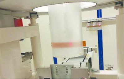
This study relates to the INFN project SYRMA‐3D for in vivo phase‐contrast breast computed tomography using the SYRMEP synchrotron radiation beamline at the ELETTRA facility in Trieste, Italy. This peculiar imaging technique uses a novel dosimetric approach with respect to the standard clinical procedure. In this study, optimization of the acquisition procedure was evaluated in terms of dose delivered to the breast. An offline dose monitoring method was also investigated using radiochromic film dosimetry. Various irradiation geometries have been investigated for scanning the prone patient's pendant breast, simulated by a 14 cm‐diameter polymethylmethacrylate cylindrical phantom containing pieces of calibrated radiochromic film type XR‐QA2. Films were inserted mid‐plane in the phantom, as well as wrapped around its external surface, and irradiated at 38 keV, with an air kerma value that would produce an estimated mean glandular dose of 5 mGy for a 14 cm‐diameter 50% glandular breast. Axial scans were performed over a full rotation or over 180°. The results point out that a scheme adopting a stepped rotation irradiation represents the best geometry to optimize the dose distribution to the breast. The feasibility of using a piece of calibrated radiochromic film wrapped around a suitable holder around the breast to monitor the scan dose offline is demonstrated.
中文翻译:

同步加速器辐射乳腺计算机断层扫描中的放射变色胶片剂量测定:一项模型研究。
这项研究与 INFN 项目 SYRMA-3D 相关,该项目使用意大利的里雅斯特 ELETTRA 设施的 SYRMEP 同步辐射束线进行体内相衬乳腺计算机断层扫描。这种独特的成像技术采用了一种符合标准临床程序的新型剂量测定方法。在这项研究中,根据传递到乳房的剂量来评估采集程序的优化。还使用放射变色胶片剂量测定法研究了离线剂量监测方法。已经研究了用于扫描俯卧患者下垂乳房的各种辐射几何形状,通过包含校准的 XR-QA2 型放射性变色胶片的 14 厘米直径聚甲基丙烯酸甲酯圆柱形模型进行模拟。将薄膜插入体模的中平面,并包裹在其外表面,并以 38 keV 进行照射,空气比释动能值对于 14 厘米直径的 50% 腺体会产生 5 mGy 的估计平均腺体剂量胸部。轴向扫描在整个旋转或超过 180° 范围内进行。结果指出,采用步进旋转照射的方案代表了优化乳房剂量分布的最佳几何形状。证明了使用一块缠绕在乳房周围合适支架上的校准放射变色薄膜来离线监测扫描剂量的可行性。
更新日期:2020-04-22
中文翻译:
同步加速器辐射乳腺计算机断层扫描中的放射变色胶片剂量测定:一项模型研究。
这项研究与 INFN 项目 SYRMA-3D 相关,该项目使用意大利的里雅斯特 ELETTRA 设施的 SYRMEP 同步辐射束线进行体内相衬乳腺计算机断层扫描。这种独特的成像技术采用了一种符合标准临床程序的新型剂量测定方法。在这项研究中,根据传递到乳房的剂量来评估采集程序的优化。还使用放射变色胶片剂量测定法研究了离线剂量监测方法。已经研究了用于扫描俯卧患者下垂乳房的各种辐射几何形状,通过包含校准的 XR-QA2 型放射性变色胶片的 14 厘米直径聚甲基丙烯酸甲酯圆柱形模型进行模拟。将薄膜插入体模的中平面,并包裹在其外表面,并以 38 keV 进行照射,空气比释动能值对于 14 厘米直径的 50% 腺体会产生 5 mGy 的估计平均腺体剂量胸部。轴向扫描在整个旋转或超过 180° 范围内进行。结果指出,采用步进旋转照射的方案代表了优化乳房剂量分布的最佳几何形状。证明了使用一块缠绕在乳房周围合适支架上的校准放射变色薄膜来离线监测扫描剂量的可行性。


 京公网安备 11010802027423号