当前位置:
X-MOL 学术
›
Pigment Cell Melanoma Res.
›
论文详情
Our official English website, www.x-mol.net, welcomes your
feedback! (Note: you will need to create a separate account there.)
Cover Image
Pigment Cell & Melanoma Research ( IF 3.9 ) Pub Date : 2020-04-22 , DOI: 10.1111/pcmr.12881
Pigment Cell & Melanoma Research ( IF 3.9 ) Pub Date : 2020-04-22 , DOI: 10.1111/pcmr.12881
|
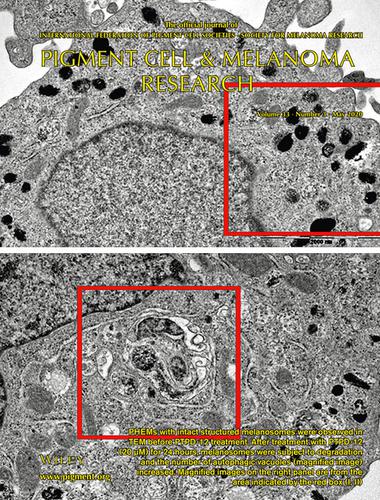
PHEMs with intact structured melanosomes were observed in TEM before PTPD‐12 treatment. After treatment with PTPD‐12 (20 μM) for 24 hours, melanosomes were subject to degradation and the number of autophagic vacuoles (magnified image) increased. Magnified images on the right panel are from the area indicated by the red box (I, II)

中文翻译:

封面图片
在PTPD-12处理之前,在TEM中观察到具有完整结构的黑色素体的PHEM。用PTPD-12(20μM)处理24小时后,黑素体遭到降解,自噬泡的数量(放大的图像)增加。右侧面板上的放大图像来自红色框(I,II)指示的区域
更新日期:2020-04-22
中文翻译:
封面图片
在PTPD-12处理之前,在TEM中观察到具有完整结构的黑色素体的PHEM。用PTPD-12(20μM)处理24小时后,黑素体遭到降解,自噬泡的数量(放大的图像)增加。右侧面板上的放大图像来自红色框(I,II)指示的区域


 京公网安备 11010802027423号