当前位置:
X-MOL 学术
›
Nat. Nanotechnol.
›
论文详情
Our official English website, www.x-mol.net, welcomes your
feedback! (Note: you will need to create a separate account there.)
Three-dimensional localization microscopy in live flowing cells.
Nature Nanotechnology ( IF 38.1 ) Pub Date : 2020-04-20 , DOI: 10.1038/s41565-020-0662-0 Lucien E Weiss 1 , Yael Shalev Ezra 1 , Sarah Goldberg 1 , Boris Ferdman 1, 2 , Omer Adir 2, 3 , Avi Schroeder 3 , Onit Alalouf 1 , Yoav Shechtman 1
Nature Nanotechnology ( IF 38.1 ) Pub Date : 2020-04-20 , DOI: 10.1038/s41565-020-0662-0 Lucien E Weiss 1 , Yael Shalev Ezra 1 , Sarah Goldberg 1 , Boris Ferdman 1, 2 , Omer Adir 2, 3 , Avi Schroeder 3 , Onit Alalouf 1 , Yoav Shechtman 1
Affiliation
|
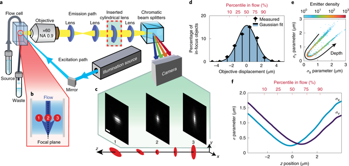
Capturing the dynamics of live cell populations with nanoscale resolution poses a significant challenge, primarily owing to the speed-resolution trade-off of existing microscopy techniques. Flow cytometry would offer sufficient throughput, but lacks subsample detail. Here we show that imaging flow cytometry, in which the point detectors of flow cytometry are replaced with a camera to record 2D images, is compatible with 3D localization microscopy through point-spread-function engineering, which encodes the depth of the emitter into the emission pattern captured by the camera. The extraction of 3D positions from sub-cellular objects of interest is achieved by calibrating the depth-dependent response of the imaging system using fluorescent beads mixed with the sample buffer. This approach enables 4D imaging of up to tens of thousands of objects per minute and can be applied to characterize chromatin dynamics and the uptake and spatial distribution of nanoparticles in live cancer cells.
中文翻译:

活细胞中的三维定位显微镜。
捕获具有纳米级分辨率的活细胞群体的动力学构成了巨大的挑战,这主要是由于现有显微镜技术在速度分辨率上的取舍。流式细胞术将提供足够的通量,但缺少子样本细节。在这里,我们显示了成像流式细胞仪,其中用相机代替流式细胞仪的点检测器来记录2D图像,通过点扩散功能工程与3D定位显微镜兼容,该技术对发射器进入发射的深度进行编码相机捕获的图案。通过使用与样品缓冲液混合的荧光珠校准成像系统的深度相关响应,可以从感兴趣的亚细胞对象中提取3D位置。
更新日期:2020-04-24
中文翻译:
活细胞中的三维定位显微镜。
捕获具有纳米级分辨率的活细胞群体的动力学构成了巨大的挑战,这主要是由于现有显微镜技术在速度分辨率上的取舍。流式细胞术将提供足够的通量,但缺少子样本细节。在这里,我们显示了成像流式细胞仪,其中用相机代替流式细胞仪的点检测器来记录2D图像,通过点扩散功能工程与3D定位显微镜兼容,该技术对发射器进入发射的深度进行编码相机捕获的图案。通过使用与样品缓冲液混合的荧光珠校准成像系统的深度相关响应,可以从感兴趣的亚细胞对象中提取3D位置。


 京公网安备 11010802027423号