当前位置:
X-MOL 学术
›
Microsc. Res. Tech.
›
论文详情
Our official English website, www.x-mol.net, welcomes your
feedback! (Note: you will need to create a separate account there.)
Observations on the oral organelle, cytopharynx, and subpellicular structure of a Dileptus sp.
Microscopy Research and Technique ( IF 2.0 ) Pub Date : 2020-04-18 , DOI: 10.1002/jemt.23498 Wenwei Liang 1, 2 , Mingjie Wang 1 , Zijian Qiu 2
Microscopy Research and Technique ( IF 2.0 ) Pub Date : 2020-04-18 , DOI: 10.1002/jemt.23498 Wenwei Liang 1, 2 , Mingjie Wang 1 , Zijian Qiu 2
Affiliation
|
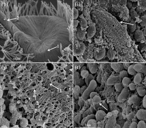
We used scanning electron microscopy (SEM) and transmission electron microscopy (TEM) to observe the oral organelle, cytopharynx, and subpellicular structure of a Dileptus sp. The main results were as follows: (a) the cytostome was located on the ventral surface of the base of the beak, surrounded by a periportal matrix that integrated 135 microtube bundles. When these microtube bundles contract, radially arranged into a disk, the cytostome was closed. When these microtube bundles were stretch, they fell into the cytostome and opens. The diameter of the cytostome was about 16 μm regardless of its closure or opening, indicating that the contraction or elongation of these microtube bundles did not change the size of the cytostome, which was only related to whether it blocked the cytostome, thus determining the opening and closing of the cytostome. There were many microtube bundles on two sides of the feeding trough, which could widen or narrow the feeding trough and facilitate beak feeding. (b) The cytopharynx was basket‐like without a bottom with a diameter of about 6 μm and was woven from two kind fibers about 0.08 and 0.19 μm. (c) There were two types of extrusomes under the pellicle. Using transmission electron microscopy,the Type I extrusomes showed narrow and long egg shape, its cross section was circular which is composed by various electronic density of concentric. Using the scanning electron microscope, they were two slightly thin clavate, the length was about 5 μm, the diameter of the middle section was about 0.75 μm, and the diameter of the two ends was about 0.32 μm, they were distributed abundantly between the microtubule fasciculi which were located on both sides of the gap on the feeding groove. Using transmission electron microscopy, the Type II extrusomes showed egg shape. Using the scanning electron microscopy, they were about 1.6 × 0.8 μm in size, they were distributed abundantly under the body pellicle while rarely the proboscis. In addition, many different of developmental stages two types of extrusomes could be also seen in the cytoplasm. (d) There were very well‐developed fibrous systems under the pellicle that were woven from fibers about 0.14 μm in diameter that attached to the pellicle and bound some organelles in the cytoplasm (e.g., mitochondria, extrusomes) and other structures to the cytoplasm and maintained cell morphology. The results of this study not only supplement and enrich the morphological contents of the Dileptus sp., but also provide the basis for the study of the taxonomy of the Dileptus sp. It also provides a new method for researchers to explore the morphology and structure of ciliate cells under the cortex by SEM.
中文翻译:

观察Dileptus sp。的口腔细胞器,细胞咽和膜下结构。
我们使用扫描电子显微镜(SEM)和透射电子显微镜(TEM)观察口服细胞器,cytopharynx,和一个的subpellicular结构Dileptussp。主要结果如下:(a)细胞体位于喙基部的腹面,周围是结合了135个微管束的门口基质。当这些微管束收缩并径向排列成圆盘时,细胞基质被封闭。当这些微管束被拉伸时,它们掉入细胞骨架并打开。不论其关闭或打开,细胞器的直径约为16μm,这表明这些微管束的收缩或伸长不会改变细胞器的大小,这仅与是否阻止细胞器有关,从而确定了开口和关闭的细胞。喂食槽的两侧有许多微管束,可以使喂食槽变宽或变窄,并有利于喙喂食。(b)细胞咽部呈篮状,无底,直径约6μm,由两种约0.08和0.19μm的纤维编织而成。(c)防护膜下有两种类型的挤出物。用透射电子显微镜观察,I型挤出物呈窄而长的卵形,其横截面为圆形,由同心的各种电子密度组成。使用扫描电子显微镜,它们是两个稍薄的棒状,长约5μm,中间部分的直径约0.75μm,两端的直径约0.32μm,它们在微管之间大量分布fasciculi位于进料槽的间隙两侧。使用透射电子显微镜观察,II型挤出物呈卵形。使用扫描电子显微镜 它们的大小约为1.6×0.8μm,分布在身体的表皮下,而长鼻很少。另外,在细胞质中还可以看到许多不同的发育阶段两种类型的挤出体。(d)防护膜下有非常发达的纤维系统,这些纤维系统由直径约0.14μm的纤维编织而成,该纤维附着在防护膜上并结合了细胞质中的一些细胞器(例如线粒体,挤压物)以及其他与细胞质和保持细胞形态。这项研究的结果不仅补充和丰富了番茄的形态学内容。(d)防护膜下有非常发达的纤维系统,这些纤维系统由直径约0.14μm的纤维编织而成,该纤维附着在防护膜上并结合了细胞质中的一些细胞器(例如线粒体,挤压物)以及其他与细胞质和保持细胞形态。这项研究的结果不仅补充和丰富了番茄的形态学内容。(d)防护膜下有非常发达的纤维系统,该系统由直径约0.14μm的纤维编织而成,该纤维附着在防护膜上并结合了细胞质中的一些细胞器(例如线粒体,挤压体)以及其他与细胞质和保持细胞形态。这项研究的结果不仅补充和丰富了番茄的形态学内容。Dileptus sp。,但也为研究Dileptus sp。的分类学提供了基础。这也为研究人员通过SEM探索皮层下纤毛细胞的形态和结构提供了新的方法。
更新日期:2020-04-18
中文翻译:
观察Dileptus sp。的口腔细胞器,细胞咽和膜下结构。
我们使用扫描电子显微镜(SEM)和透射电子显微镜(TEM)观察口服细胞器,cytopharynx,和一个的subpellicular结构Dileptussp。主要结果如下:(a)细胞体位于喙基部的腹面,周围是结合了135个微管束的门口基质。当这些微管束收缩并径向排列成圆盘时,细胞基质被封闭。当这些微管束被拉伸时,它们掉入细胞骨架并打开。不论其关闭或打开,细胞器的直径约为16μm,这表明这些微管束的收缩或伸长不会改变细胞器的大小,这仅与是否阻止细胞器有关,从而确定了开口和关闭的细胞。喂食槽的两侧有许多微管束,可以使喂食槽变宽或变窄,并有利于喙喂食。(b)细胞咽部呈篮状,无底,直径约6μm,由两种约0.08和0.19μm的纤维编织而成。(c)防护膜下有两种类型的挤出物。用透射电子显微镜观察,I型挤出物呈窄而长的卵形,其横截面为圆形,由同心的各种电子密度组成。使用扫描电子显微镜,它们是两个稍薄的棒状,长约5μm,中间部分的直径约0.75μm,两端的直径约0.32μm,它们在微管之间大量分布fasciculi位于进料槽的间隙两侧。使用透射电子显微镜观察,II型挤出物呈卵形。使用扫描电子显微镜 它们的大小约为1.6×0.8μm,分布在身体的表皮下,而长鼻很少。另外,在细胞质中还可以看到许多不同的发育阶段两种类型的挤出体。(d)防护膜下有非常发达的纤维系统,这些纤维系统由直径约0.14μm的纤维编织而成,该纤维附着在防护膜上并结合了细胞质中的一些细胞器(例如线粒体,挤压物)以及其他与细胞质和保持细胞形态。这项研究的结果不仅补充和丰富了番茄的形态学内容。(d)防护膜下有非常发达的纤维系统,这些纤维系统由直径约0.14μm的纤维编织而成,该纤维附着在防护膜上并结合了细胞质中的一些细胞器(例如线粒体,挤压物)以及其他与细胞质和保持细胞形态。这项研究的结果不仅补充和丰富了番茄的形态学内容。(d)防护膜下有非常发达的纤维系统,该系统由直径约0.14μm的纤维编织而成,该纤维附着在防护膜上并结合了细胞质中的一些细胞器(例如线粒体,挤压体)以及其他与细胞质和保持细胞形态。这项研究的结果不仅补充和丰富了番茄的形态学内容。Dileptus sp。,但也为研究Dileptus sp。的分类学提供了基础。这也为研究人员通过SEM探索皮层下纤毛细胞的形态和结构提供了新的方法。


 京公网安备 11010802027423号