当前位置:
X-MOL 学术
›
Cytom. Part A
›
论文详情
Our official English website, www.x-mol.net, welcomes your feedback! (Note: you will need to create a separate account there.)
Coregistered Spectral Optical Coherence Tomography and Two-Photon Microscopy for Multimodal Near-Instantaneous Deep-Tissue Imaging.
Cytometry Part A ( IF 3.7 ) Pub Date : 2020-04-15 , DOI: 10.1002/cyto.a.24012 Asylkhan Rakhymzhan 1 , Lucie Reuter 1 , Raphael Raspe 2, 3 , Daniel Bremer 1 , Robert Günther 1, 2 , Ruth Leben 1 , Judith Heidelin 4 , Volker Andresen 4 , Sergey Cheremukhin 4 , Hinnerk Schulz-Hildebrandt 5 , Maria G Bixel 6 , Ralf H Adams 6 , Helena Radbruch 7 , Gereon Hüttmann 5, 8 , Anja E Hauser 2, 3 , Raluca A Niesner 1, 9
Cytometry Part A ( IF 3.7 ) Pub Date : 2020-04-15 , DOI: 10.1002/cyto.a.24012 Asylkhan Rakhymzhan 1 , Lucie Reuter 1 , Raphael Raspe 2, 3 , Daniel Bremer 1 , Robert Günther 1, 2 , Ruth Leben 1 , Judith Heidelin 4 , Volker Andresen 4 , Sergey Cheremukhin 4 , Hinnerk Schulz-Hildebrandt 5 , Maria G Bixel 6 , Ralf H Adams 6 , Helena Radbruch 7 , Gereon Hüttmann 5, 8 , Anja E Hauser 2, 3 , Raluca A Niesner 1, 9
Affiliation
|
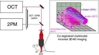
Two-photon microscopy (2PM) has brought unique insight into the mechanisms underlying immune system dynamics and function since it enables monitoring of cellular motility and communication in complex systems within their genuine environment-the living organism. However, use of 2PM in clinical settings is limited. In contrast, optical coherence tomography (OCT), a noninvasive label-free diagnostic imaging method, which allows monitoring morphologic changes of large tissue regions in vivo, has found broad application in the clinic. Here we developed a combined multimodal technology to achieve near-instantaneous coregistered OCT, 2PM, and second harmonic generation (SHG) imaging over large volumes (up to 1,000 × 1,000 × 300 μm3 ) of tendons and other tissue compartments in mouse paws, as well as in mouse lymph nodes, spleens, and femurs. Using our multimodal imaging approach, we found differences in macrophage cell shape and motility behavior depending on whether they are located in tendons or in the surrounding tissue compartments of the mouse paw. The cellular shape of tissue-resident macrophages, indicative for their role in tissue, correlated with the supramolecular organization of collagen as revealed by SHG and OCT. Hence, the here-presented approach of coregistered OCT and 2PM has the potential to link specific cellular phenotypes and functions (as revealed by 2PM) to tissue morphology (as highlighted by OCT) and thus, to build a bridge between basic research knowledge and clinical observations. © 2020 The Authors. Cytometry Part A published by Wiley Periodicals, Inc. on behalf of International Society for Advancement of Cytometry.
中文翻译:

用于多模态近瞬时深部组织成像的配准光谱光学相干断层扫描和双光子显微镜。
双光子显微镜 (2PM) 为免疫系统动力学和功能的潜在机制带来了独特的见解,因为它能够监测真实环境——活生物体——复杂系统中的细胞运动和通信。然而,在临床环境中使用 2PM 是有限的。相比之下,光学相干断层扫描 (OCT) 是一种无创无标记诊断成像方法,可以监测体内大组织区域的形态变化,已在临床中得到广泛应用。在这里,我们开发了一种组合多模态技术,以实现对大体积(高达 1,000 × 1,000 × 300 μm3)的肌腱和鼠标爪中的其他组织隔室的近乎瞬时配准的 OCT、2PM 和二次谐波生成 (SHG) 成像如小鼠淋巴结、脾脏和股骨。使用我们的多模态成像方法,我们发现巨噬细胞形状和运动行为的差异取决于它们是位于肌腱中还是位于小鼠爪子的周围组织隔室中。组织驻留巨噬细胞的细胞形状表明它们在组织中的作用,与 SHG 和 OCT 揭示的胶原蛋白的超分子组织相关。因此,这里介绍的联合 OCT 和 2PM 的方法有可能将特定的细胞表型和功能(如 2PM 所揭示的)与组织形态学(如 OCT 所强调的)联系起来,从而在基础研究知识和临床之间架起一座桥梁。观察。© 2020 作者。Cytometry Part A 由 Wiley Periodicals, Inc. 代表 International Society for Advancement of Cytometry 出版。我们发现巨噬细胞形状和运动行为的差异取决于它们是位于肌腱中还是位于小鼠爪子的周围组织隔室中。SHG 和 OCT 揭示的组织驻留巨噬细胞的细胞形状,表明它们在组织中的作用,与胶原蛋白的超分子组织相关。因此,这里介绍的联合 OCT 和 2PM 的方法有可能将特定的细胞表型和功能(如 2PM 所揭示的)与组织形态学(如 OCT 所强调的)联系起来,从而在基础研究知识和临床研究之间架起一座桥梁。观察。© 2020 作者。Cytometry Part A 由 Wiley Periodicals, Inc. 代表 International Society for Advancement of Cytometry 出版。我们发现巨噬细胞形状和运动行为的差异取决于它们是位于肌腱中还是位于小鼠爪子的周围组织隔室中。SHG 和 OCT 揭示的组织驻留巨噬细胞的细胞形状,表明它们在组织中的作用,与胶原蛋白的超分子组织相关。因此,这里介绍的联合 OCT 和 2PM 的方法有可能将特定的细胞表型和功能(如 2PM 所揭示的)与组织形态学(如 OCT 所强调的)联系起来,从而在基础研究知识和临床之间架起一座桥梁。观察。© 2020 作者。Cytometry Part A 由 Wiley Periodicals, Inc. 代表 International Society for Advancement of Cytometry 出版。
更新日期:2020-04-15
中文翻译:
用于多模态近瞬时深部组织成像的配准光谱光学相干断层扫描和双光子显微镜。
双光子显微镜 (2PM) 为免疫系统动力学和功能的潜在机制带来了独特的见解,因为它能够监测真实环境——活生物体——复杂系统中的细胞运动和通信。然而,在临床环境中使用 2PM 是有限的。相比之下,光学相干断层扫描 (OCT) 是一种无创无标记诊断成像方法,可以监测体内大组织区域的形态变化,已在临床中得到广泛应用。在这里,我们开发了一种组合多模态技术,以实现对大体积(高达 1,000 × 1,000 × 300 μm3)的肌腱和鼠标爪中的其他组织隔室的近乎瞬时配准的 OCT、2PM 和二次谐波生成 (SHG) 成像如小鼠淋巴结、脾脏和股骨。使用我们的多模态成像方法,我们发现巨噬细胞形状和运动行为的差异取决于它们是位于肌腱中还是位于小鼠爪子的周围组织隔室中。组织驻留巨噬细胞的细胞形状表明它们在组织中的作用,与 SHG 和 OCT 揭示的胶原蛋白的超分子组织相关。因此,这里介绍的联合 OCT 和 2PM 的方法有可能将特定的细胞表型和功能(如 2PM 所揭示的)与组织形态学(如 OCT 所强调的)联系起来,从而在基础研究知识和临床之间架起一座桥梁。观察。© 2020 作者。Cytometry Part A 由 Wiley Periodicals, Inc. 代表 International Society for Advancement of Cytometry 出版。我们发现巨噬细胞形状和运动行为的差异取决于它们是位于肌腱中还是位于小鼠爪子的周围组织隔室中。SHG 和 OCT 揭示的组织驻留巨噬细胞的细胞形状,表明它们在组织中的作用,与胶原蛋白的超分子组织相关。因此,这里介绍的联合 OCT 和 2PM 的方法有可能将特定的细胞表型和功能(如 2PM 所揭示的)与组织形态学(如 OCT 所强调的)联系起来,从而在基础研究知识和临床研究之间架起一座桥梁。观察。© 2020 作者。Cytometry Part A 由 Wiley Periodicals, Inc. 代表 International Society for Advancement of Cytometry 出版。我们发现巨噬细胞形状和运动行为的差异取决于它们是位于肌腱中还是位于小鼠爪子的周围组织隔室中。SHG 和 OCT 揭示的组织驻留巨噬细胞的细胞形状,表明它们在组织中的作用,与胶原蛋白的超分子组织相关。因此,这里介绍的联合 OCT 和 2PM 的方法有可能将特定的细胞表型和功能(如 2PM 所揭示的)与组织形态学(如 OCT 所强调的)联系起来,从而在基础研究知识和临床之间架起一座桥梁。观察。© 2020 作者。Cytometry Part A 由 Wiley Periodicals, Inc. 代表 International Society for Advancement of Cytometry 出版。


 京公网安备 11010802027423号