当前位置:
X-MOL 学术
›
J. Raman Spectrosc.
›
论文详情
Our official English website, www.x-mol.net, welcomes your
feedback! (Note: you will need to create a separate account there.)
Raman spectroscopic analysis and imaging in two cases of benign cementoma: Comparison with dental and bone tissues
Journal of Raman Spectroscopy ( IF 2.4 ) Pub Date : 2020-04-14 , DOI: 10.1002/jrs.5880 Daniel Chappard 1 , Bernard Guillaume 1, 2 , Gil Teman 3 , Jean‐Daniel Kün‐Darbois 1, 4
Journal of Raman Spectroscopy ( IF 2.4 ) Pub Date : 2020-04-14 , DOI: 10.1002/jrs.5880 Daniel Chappard 1 , Bernard Guillaume 1, 2 , Gil Teman 3 , Jean‐Daniel Kün‐Darbois 1, 4
Affiliation
|
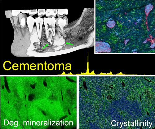
Benign cementoma is a rare tumor of the jaws whose origin is still debating. The tumor may derive from the dental cementum or the bone surrounding the socket. We have studied the profiles of enamel, dentine, and cementum from normal teeth and alveolar and cortical bone of the mandible. Raman spectroscopy was used to compare the spectra of these tissues embedded in poly(methylmethacrylate) from 300 to 3,100 cm−1. The degree of mineralization, carbonate‐to‐phosphate ratio, and crystallinity were derived. Tissue hydration was determined by measuring the ratio of the 813 cm−1 band of methacrylate to the 960 cm−1 band of phosphate. Raman spectroscopic imaging was obtained on large tissue areas with a bandwidth from 500 to 1,620 cm−1. Maps of the different peak ratio were obtained to analyze the mineral and organic phases (crystallinity, carbonate‐to‐phosphate, degree of mineralization) together with tissue hydration. Cementomas appeared highly heterogeneous, and their matrix possessed histological and spectroscopic characteristics that resemble highly calcified woven bone with a poor organization of the hydroxyapatite crystals.
中文翻译:

拉曼光谱分析和影像学检查在两例良性牙骨瘤中:与牙齿和骨组织的比较
良性牙骨质瘤是一种罕见的颌骨肿瘤,其起源仍在争论中。肿瘤可源自牙骨质或窝周围的骨头。我们研究了正常牙齿,下颌骨的牙槽和皮质骨的牙釉质,牙本质和牙骨质的轮廓。拉曼光谱法用于比较嵌入到聚甲基丙烯酸甲酯中的这些组织的300至3,100 cm -1的光谱。得出矿化度,碳酸盐与磷酸盐之比和结晶度。组织水合,通过测量的813厘米的比率来确定-1甲基丙烯酸酯的频带到960厘米-1磷酸盐的频带。拉曼光谱成像在带宽为500至1,620 cm -1的大组织区域上获得。获得了不同峰比率的图,以分析矿物和有机相(结晶度,碳酸盐对磷酸盐,矿化度)以及组织的水合作用。牙骨质瘤高度异质,其基质具有组织学和光谱学特征,类似于高度钙化的编织骨,羟基磷灰石晶体的组织较差。
更新日期:2020-04-14
中文翻译:
拉曼光谱分析和影像学检查在两例良性牙骨瘤中:与牙齿和骨组织的比较
良性牙骨质瘤是一种罕见的颌骨肿瘤,其起源仍在争论中。肿瘤可源自牙骨质或窝周围的骨头。我们研究了正常牙齿,下颌骨的牙槽和皮质骨的牙釉质,牙本质和牙骨质的轮廓。拉曼光谱法用于比较嵌入到聚甲基丙烯酸甲酯中的这些组织的300至3,100 cm -1的光谱。得出矿化度,碳酸盐与磷酸盐之比和结晶度。组织水合,通过测量的813厘米的比率来确定-1甲基丙烯酸酯的频带到960厘米-1磷酸盐的频带。拉曼光谱成像在带宽为500至1,620 cm -1的大组织区域上获得。获得了不同峰比率的图,以分析矿物和有机相(结晶度,碳酸盐对磷酸盐,矿化度)以及组织的水合作用。牙骨质瘤高度异质,其基质具有组织学和光谱学特征,类似于高度钙化的编织骨,羟基磷灰石晶体的组织较差。


 京公网安备 11010802027423号