当前位置:
X-MOL 学术
›
Eur. J. Neurosci.
›
论文详情
Our official English website, www.x-mol.net, welcomes your
feedback! (Note: you will need to create a separate account there.)
Maturation of the microglial population varies across mesolimbic nuclei.
European Journal of Neuroscience ( IF 2.7 ) Pub Date : 2020-04-13 , DOI: 10.1111/ejn.14740 Keenan T Hope 1 , Isobel A Hawes 2 , Eric N Moca 1 , Antonello Bonci , Lindsay M De Biase 1, 2
European Journal of Neuroscience ( IF 2.7 ) Pub Date : 2020-04-13 , DOI: 10.1111/ejn.14740 Keenan T Hope 1 , Isobel A Hawes 2 , Eric N Moca 1 , Antonello Bonci , Lindsay M De Biase 1, 2
Affiliation
|
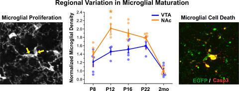
Microglia play critical roles during CNS development and undergo dramatic changes in tissue distribution, morphology, and gene expression as they transition from embryonic to neonatal to adult microglial phenotypes. Despite the magnitude of these phenotypic shifts, little is known about the time course and dynamics of these transitions and whether they vary across brain regions. Here, we define the time course of microglial maturation in key regions of the basal ganglia in mice, where significant regional differences in microglial phenotype are present in adults. We found that microglial density peaks in the ventral tegmental area (VTA) and nucleus accumbens (NAc) during the third postnatal week, driven by a burst of microglial proliferation. Microglial abundance is then refined to adult levels through a combination of tissue expansion and microglial programmed cell death. This overproduction and refinement of microglia was significantly more pronounced in the NAc than in the VTA and was accompanied by a sharp peak in NAc microglial lysosome abundance in the third postnatal week. Collectively, these data identify a key developmental window when elevated microglial density in discrete basal ganglia nuclei may support circuit refinement and could increase susceptibility to inflammatory insults.
中文翻译:

小胶质细胞群体的成熟在中脑边缘核之间变化。
小胶质细胞在中枢神经系统的发育过程中起着至关重要的作用,并且在它们从胚胎到新生儿再到成年的小胶质细胞表型转变时,它们在组织分布,形态和基因表达方面发生了巨大变化。尽管这些表型变化的幅度很大,但对于这些跃迁的时程和动态以及它们是否在整个大脑区域变化都知之甚少。在这里,我们定义了小鼠基底神经节关键区域小胶质细胞成熟的时间过程,其中成年小鼠存在小胶质细胞表型的显着区域差异。我们发现,在出生后的第三周,小胶质细胞增殖的爆发驱动了腹侧被盖区(VTA)和伏隔核(NAc)中的小胶质细胞密度峰值。然后,通过组织扩张和小胶质细胞程序性死亡的组合,将小胶质细胞的丰度提高到成年水平。在NAc中,这种小胶质细胞的过度生产和细化比在VTA中更为明显,并且在产后第三周伴随着NAc小胶质细胞溶酶体丰度的急剧增加。总体而言,这些数据确定了离散的基底神经节核中的小胶质细胞密度升高可能支持回路细化并可能增加对炎症性损伤的敏感性时,确定了关键的发育窗口。
更新日期:2020-05-11
中文翻译:
小胶质细胞群体的成熟在中脑边缘核之间变化。
小胶质细胞在中枢神经系统的发育过程中起着至关重要的作用,并且在它们从胚胎到新生儿再到成年的小胶质细胞表型转变时,它们在组织分布,形态和基因表达方面发生了巨大变化。尽管这些表型变化的幅度很大,但对于这些跃迁的时程和动态以及它们是否在整个大脑区域变化都知之甚少。在这里,我们定义了小鼠基底神经节关键区域小胶质细胞成熟的时间过程,其中成年小鼠存在小胶质细胞表型的显着区域差异。我们发现,在出生后的第三周,小胶质细胞增殖的爆发驱动了腹侧被盖区(VTA)和伏隔核(NAc)中的小胶质细胞密度峰值。然后,通过组织扩张和小胶质细胞程序性死亡的组合,将小胶质细胞的丰度提高到成年水平。在NAc中,这种小胶质细胞的过度生产和细化比在VTA中更为明显,并且在产后第三周伴随着NAc小胶质细胞溶酶体丰度的急剧增加。总体而言,这些数据确定了离散的基底神经节核中的小胶质细胞密度升高可能支持回路细化并可能增加对炎症性损伤的敏感性时,确定了关键的发育窗口。


 京公网安备 11010802027423号