当前位置:
X-MOL 学术
›
J. Synchrotron Radiat.
›
论文详情
Our official English website, www.x-mol.net, welcomes your
feedback! (Note: you will need to create a separate account there.)
LamNI - an instrument for X-ray scanning microscopy in laminography geometry.
Journal of Synchrotron Radiation ( IF 2.4 ) Pub Date : 2020-04-06 , DOI: 10.1107/s1600577520003586 Mirko Holler 1 , Michal Odstrčil 1 , Manuel Guizar-Sicairos 1 , Maxime Lebugle 1 , Ulrich Frommherz 2 , Thierry Lachat 3 , Oliver Bunk 1 , Joerg Raabe 1 , Gabriel Aeppli 1
Journal of Synchrotron Radiation ( IF 2.4 ) Pub Date : 2020-04-06 , DOI: 10.1107/s1600577520003586 Mirko Holler 1 , Michal Odstrčil 1 , Manuel Guizar-Sicairos 1 , Maxime Lebugle 1 , Ulrich Frommherz 2 , Thierry Lachat 3 , Oliver Bunk 1 , Joerg Raabe 1 , Gabriel Aeppli 1
Affiliation
|
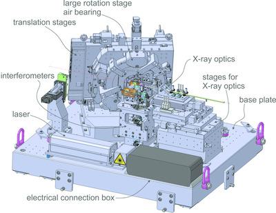
Across all branches of science, medicine and engineering, high-resolution microscopy is required to understand functionality. Although optical methods have been developed to `defeat' the diffraction limit and produce 3D images, and electrons have proven ever more useful in creating pictures of small objects or thin sections, so far there is no substitute for X-ray microscopy in providing multiscale 3D images of objects with a single instrument and minimal labeling and preparation. A powerful technique proven to continuously access length scales from 10 nm to 10 µm is ptychographic X-ray computed tomography, which, on account of the orthogonality of the tomographic rotation axis to the illuminating beam, still has the limitation of necessitating pillar-shaped samples of small (ca 10 µm) diameter. Large-area planar samples are common in science and engineering, and it is therefore highly desirable to create an X-ray microscope that can examine such samples without the extraction of pillars. Computed laminography, where the axis of rotation is not perpendicular to the illumination direction, solves this problem. This entailed the development of a new instrument, LamNI, dedicated to high-resolution 3D scanning X-ray microscopy via hard X-ray ptychographic laminography. Scanning precision is achieved by a dedicated interferometry scheme and the instrument covers a scan range of 12 mm × 12 mm with a position stability of 2 nm and positioning errors below 5 nm. A new feature of LamNI is a pair of counter-rotating stages carrying the sample and interferometric mirrors, respectively.
中文翻译:

LamNI - 层层摄影几何中的 X 射线扫描显微镜仪器。
在科学、医学和工程的所有分支中,都需要高分辨率显微镜来了解功能。尽管光学方法已经被开发出来,可以“打破”衍射极限并产生 3D 图像,并且电子已被证明在创建小物体或薄片的图像方面更加有用,但到目前为止,在提供多尺度 3D 图像方面,X 射线显微镜无法替代使用单一仪器和最少的标记和准备即可获得物体图像。叠层 X 射线计算机断层扫描是一种被证明可以连续获取 10 nm 至 10 µm 长度尺度的强大技术,但由于断层扫描旋转轴与照明光束的正交性,该技术仍然存在需要柱状样品的局限性小直径(约 10 µm)。大面积平面样品在科学和工程中很常见,因此非常需要创建一种无需提取支柱即可检查此类样品的 X 射线显微镜。旋转轴不垂直于照明方向的计算机断层摄影解决了这个问题。这就需要开发一种新仪器 LamNI,专用于通过硬 X 射线叠层摄影进行高分辨率 3D 扫描 X 射线显微镜。扫描精度通过专用干涉测量方案实现,仪器扫描范围为12 mm×12 mm,位置稳定性为2 nm,定位误差低于5 nm。LamNI 的一个新功能是一对分别承载样本和干涉镜的反向旋转平台。
更新日期:2020-04-06
中文翻译:
LamNI - 层层摄影几何中的 X 射线扫描显微镜仪器。
在科学、医学和工程的所有分支中,都需要高分辨率显微镜来了解功能。尽管光学方法已经被开发出来,可以“打破”衍射极限并产生 3D 图像,并且电子已被证明在创建小物体或薄片的图像方面更加有用,但到目前为止,在提供多尺度 3D 图像方面,X 射线显微镜无法替代使用单一仪器和最少的标记和准备即可获得物体图像。叠层 X 射线计算机断层扫描是一种被证明可以连续获取 10 nm 至 10 µm 长度尺度的强大技术,但由于断层扫描旋转轴与照明光束的正交性,该技术仍然存在需要柱状样品的局限性小直径(约 10 µm)。大面积平面样品在科学和工程中很常见,因此非常需要创建一种无需提取支柱即可检查此类样品的 X 射线显微镜。旋转轴不垂直于照明方向的计算机断层摄影解决了这个问题。这就需要开发一种新仪器 LamNI,专用于通过硬 X 射线叠层摄影进行高分辨率 3D 扫描 X 射线显微镜。扫描精度通过专用干涉测量方案实现,仪器扫描范围为12 mm×12 mm,位置稳定性为2 nm,定位误差低于5 nm。LamNI 的一个新功能是一对分别承载样本和干涉镜的反向旋转平台。


 京公网安备 11010802027423号