当前位置:
X-MOL 学术
›
J. Synchrotron Radiat.
›
论文详情
Our official English website, www.x-mol.net, welcomes your
feedback! (Note: you will need to create a separate account there.)
A single-crystal diamond X-ray pixel detector with embedded graphitic electrodes.
Journal of Synchrotron Radiation ( IF 2.4 ) Pub Date : 2020-03-31 , DOI: 10.1107/s160057752000140x C Bloomer 1 , M E Newton 1 , G Rehm 2 , P S Salter 3
Journal of Synchrotron Radiation ( IF 2.4 ) Pub Date : 2020-03-31 , DOI: 10.1107/s160057752000140x C Bloomer 1 , M E Newton 1 , G Rehm 2 , P S Salter 3
Affiliation
|
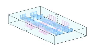
The first experimental results from a new transmissive diagnostic instrument for synchrotron X-ray beamlines are presented. The instrument utilizes a single-crystal chemical-vapour-deposition diamond plate as the detector material, with graphitic wires embedded within the bulk diamond acting as electrodes. The resulting instrument is an all-carbon transmissive X-ray imaging detector. Within the instrument's transmissive aperture there is no surface metallization that could absorb X-rays, and no surface structures that could be damaged by exposure to synchrotron X-ray beams. The graphitic electrodes are fabricated in situ within the bulk diamond using a laser-writing technique. Two separate arrays of parallel graphitic wires are fabricated, running parallel to the diamond surface and perpendicular to each other, at two different depths within the diamond. One array of wires has a modulated bias voltage applied; the perpendicular array is a series of readout electrodes. X-rays passing through the detector generate charge carriers within the bulk diamond through photoionization, and these charge carriers travel to the nearest readout electrode under the influence of the modulated electrical bias. Each of the crossing points between perpendicular wires acts as an individual pixel. The simultaneous read-out of all pixels is achieved using a lock-in technique. The parallel wires within each array are separated by 50 µm, determining the pixel pitch. Readout is obtained at 100 Hz, and the resolution of the X-ray beam position measurement is 600 nm for a 180 µm size beam.
中文翻译:

具有嵌入式石墨电极的单晶金刚石 X 射线像素探测器。
提出了同步加速器 X 射线束线的新型透射诊断仪器的第一个实验结果。该仪器采用单晶化学气相沉积金刚石板作为探测器材料,嵌入块状金刚石内的石墨线充当电极。由此产生的仪器是全碳透射式 X 射线成像探测器。在仪器的透射孔径内,没有可以吸收 X 射线的表面金属化层,也没有可能因暴露于同步加速器 X 射线束而损坏的表面结构。使用激光写入技术在块状金刚石内原位制造石墨电极。制造了两个独立的平行石墨线阵列,在金刚石内的两个不同深度处平行于金刚石表面延伸且彼此垂直。一组导线施加了调制偏置电压;垂直阵列是一系列读出电极。穿过探测器的 X 射线通过光电离在块状金刚石内产生电荷载流子,这些电荷载流子在调制电偏压的影响下行进到最近的读出电极。垂直线之间的每个交叉点都充当单独的像素。使用锁定技术可同时读出所有像素。每个阵列内的平行线间隔 50 µm,决定像素间距。读数以 100 Hz 获得,对于 180 µm 尺寸的光束,X 射线束位置测量的分辨率为 600 nm。
更新日期:2020-03-31
中文翻译:
具有嵌入式石墨电极的单晶金刚石 X 射线像素探测器。
提出了同步加速器 X 射线束线的新型透射诊断仪器的第一个实验结果。该仪器采用单晶化学气相沉积金刚石板作为探测器材料,嵌入块状金刚石内的石墨线充当电极。由此产生的仪器是全碳透射式 X 射线成像探测器。在仪器的透射孔径内,没有可以吸收 X 射线的表面金属化层,也没有可能因暴露于同步加速器 X 射线束而损坏的表面结构。使用激光写入技术在块状金刚石内原位制造石墨电极。制造了两个独立的平行石墨线阵列,在金刚石内的两个不同深度处平行于金刚石表面延伸且彼此垂直。一组导线施加了调制偏置电压;垂直阵列是一系列读出电极。穿过探测器的 X 射线通过光电离在块状金刚石内产生电荷载流子,这些电荷载流子在调制电偏压的影响下行进到最近的读出电极。垂直线之间的每个交叉点都充当单独的像素。使用锁定技术可同时读出所有像素。每个阵列内的平行线间隔 50 µm,决定像素间距。读数以 100 Hz 获得,对于 180 µm 尺寸的光束,X 射线束位置测量的分辨率为 600 nm。


 京公网安备 11010802027423号