Our official English website, www.x-mol.net, welcomes your
feedback! (Note: you will need to create a separate account there.)
Autoradiographic mapping of synaptic vesicle glycoprotein 2A in non-human primate and human brain.
SYNAPSE ( IF 1.6 ) Pub Date : 2020-04-07 , DOI: 10.1002/syn.22157 Katarina Varnäs 1 , Vladimir Stepanov 1 , Christer Halldin 1
SYNAPSE ( IF 1.6 ) Pub Date : 2020-04-07 , DOI: 10.1002/syn.22157 Katarina Varnäs 1 , Vladimir Stepanov 1 , Christer Halldin 1
Affiliation
|
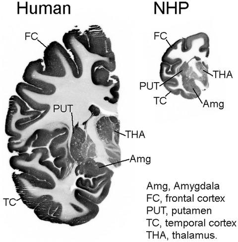
Synaptic vesicle glycoprotein 2A (SV2A) has been previously characterized as an imaging biomarker for assessment of synaptic density in positron emission tomography (PET) studies of patients with neurological conditions. To provide detailed maps of the brain localization of SV2A autoradiography studies were carried out using the SV2A radioligand [11C]UCB‐J and whole hemisphere sections of non‐human primate (NHP) and human brain. Binding of [11C]UCB‐J was observed in all evaluated grey matter structures of the primate brain, with highest density in the caudate nucleus and cortex and lowest density in pons and globus pallidus. The density of [11C]UCB‐J binding sites in human brain showed a good correlation with that in NHP brain. Binding of [11C]UCB‐J in the white matter was very low relative to that in grey matter containing structures and was only inhibited to a minor extent by co‐incubation with a saturating concentration of unlabelled UCB‐J. The high‐resolution images obtained in the present study may aid the interpretation of data acquired in human subjects examined using [11C]UCB‐J in PET studies. In addition, observation of low binding for [11C]UCB‐J in white matter (centrum semiovale) supports that this structure can be used as a reference region for quantitative analysis of [11C]UCB‐J PET data.
中文翻译:

非人类灵长类动物和人类大脑中突触囊泡糖蛋白 2A 的放射自显影图。
突触囊泡糖蛋白 2A (SV2A) 先前已被表征为用于评估神经系统疾病患者正电子发射断层扫描 (PET) 研究中突触密度的成像生物标志物。为了提供 SV2A 放射自显影研究的大脑定位的详细图谱,使用 SV2A 放射性配体 [ 11 C] UCB-J 和非人类灵长类动物 (NHP) 和人类大脑的整个半球切片进行了研究。在所有评估的灵长类大脑灰质结构中都观察到[ 11 C]UCB-J 的结合,尾状核和皮层的密度最高,脑桥和苍白球的密度最低。人脑中[ 11 C]UCB-J 结合位点的密度与 NHP 脑中的相关性良好。[ 11 的绑定白质中的 C]UCB-J 相对于包含灰质的结构而言非常低,并且仅通过与饱和浓度的未标记 UCB-J 共同孵育而在较小程度上受到抑制。本研究中获得的高分辨率图像可能有助于解释在 PET 研究中使用 [ 11 C] UCB-J检查的人类受试者中获得的数据。此外,在白质(半卵圆中心)中观察到 [ 11 C]UCB-J的低结合支持该结构可用作 [ 11 C]UCB-J PET 数据定量分析的参考区域。
更新日期:2020-04-07
中文翻译:
非人类灵长类动物和人类大脑中突触囊泡糖蛋白 2A 的放射自显影图。
突触囊泡糖蛋白 2A (SV2A) 先前已被表征为用于评估神经系统疾病患者正电子发射断层扫描 (PET) 研究中突触密度的成像生物标志物。为了提供 SV2A 放射自显影研究的大脑定位的详细图谱,使用 SV2A 放射性配体 [ 11 C] UCB-J 和非人类灵长类动物 (NHP) 和人类大脑的整个半球切片进行了研究。在所有评估的灵长类大脑灰质结构中都观察到[ 11 C]UCB-J 的结合,尾状核和皮层的密度最高,脑桥和苍白球的密度最低。人脑中[ 11 C]UCB-J 结合位点的密度与 NHP 脑中的相关性良好。[ 11 的绑定白质中的 C]UCB-J 相对于包含灰质的结构而言非常低,并且仅通过与饱和浓度的未标记 UCB-J 共同孵育而在较小程度上受到抑制。本研究中获得的高分辨率图像可能有助于解释在 PET 研究中使用 [ 11 C] UCB-J检查的人类受试者中获得的数据。此外,在白质(半卵圆中心)中观察到 [ 11 C]UCB-J的低结合支持该结构可用作 [ 11 C]UCB-J PET 数据定量分析的参考区域。


 京公网安备 11010802027423号