当前位置:
X-MOL 学术
›
Microsc. Res. Tech.
›
论文详情
Our official English website, www.x-mol.net, welcomes your
feedback! (Note: you will need to create a separate account there.)
The structure of cortical bone as revealed by the application of methods for the calcified matrix study.
Microscopy Research and Technique ( IF 2.0 ) Pub Date : 2020-03-30 , DOI: 10.1002/jemt.23477 Ugo E Pazzaglia 1 , Marcella Reguzzoni 2 , Laura Depero 3 , Stefania Federici 3 , Mariapia Bondioni 1 , Guido Zarattini 1 , Mario Raspanti 2
Microscopy Research and Technique ( IF 2.0 ) Pub Date : 2020-03-30 , DOI: 10.1002/jemt.23477 Ugo E Pazzaglia 1 , Marcella Reguzzoni 2 , Laura Depero 3 , Stefania Federici 3 , Mariapia Bondioni 1 , Guido Zarattini 1 , Mario Raspanti 2
Affiliation
|
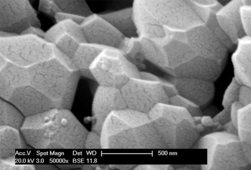
Calcination and decalcification are basic procedures useful to a morphological approach of a biological, composite material like cortical bone. The study was carried out on a whole human femur conserved in liquid (from an educational collection). Cortical fracturing and SEM observation of vascular canals surface collagen texture was used to study bone deproteination at scalar temperatures (400–1,200°C) and acid bone decalcification at crescent time intervals. Heating burned and vaporized the organic matrix with shrinkage of the bone specimens as documented by the weight loss and transverse surface morphometry. SEM showed a pattern of aligned spherulites at 400°C which maintained the collagen fibrils layout (like a mineral cast), followed by a spherulites fusion progression with the temperature increments. At 1200°C a crystalline‐like structure of tightly‐packed trapezohendron units. XRD analysis supported the SEM morphology displaying the complete Debey rings of hydroxyapatite and spotted Debey rings of withlockite. Surface Ca and P elution was documented after 12 hr of exposition to the acid solution by dissolution of spherulites and the whole canal surface decalcified in depth after 15 days by SEM‐EDAX analysis. The periodic pattern of collagen fibrils was still evident up to 15 days of decalcification together with fine granular deposits of a not‐collagenic proteic material, while after 30 days no period was observed in the decalcified fibrils.
中文翻译:

通过应用钙化基质研究方法揭示的皮质骨结构。
煅烧和脱钙是对生物复合材料(如皮质骨)的形态学方法有用的基本程序。该研究是对保存在液体中的整个人类股骨进行的(来自教育机构)。皮层破裂和血管管表面胶原蛋白的SEM观察被用来研究标量温度(400–1,200°C)下的骨脱蛋白和新月时间间隔的酸性骨脱钙。加热使有机基质燃烧和汽化,并伴随骨标本的收缩,这是通过重量损失和横向表面形貌证明的。SEM显示了在400℃下排列的球晶的图案,其维持了胶原原纤维的排列(像矿物铸模一样),随后随着温度的升高,球晶融合发展。在1200°C下,紧密堆积的梯形电子单元呈晶体状结构。XRD分析支持SEM形态,显示了完整的羟基磷灰石Debey环和点状钙石的Debey环。暴露于酸性溶液中12个小时后,通过球晶的溶解记录了表面Ca和P的溶出,并在15天后通过SEM-EDAX分析对整个运河表面进行了深度脱钙。直至15天的脱钙以及非胶原蛋白蛋白的细小颗粒沉积,胶原原纤维的周期性模式仍然很明显,而在30天后,未钙化的原纤维中没有观察到周期。暴露在酸性溶液中12个小时后,通过球晶的溶解记录了表面Ca和P的溶出,并在15天后通过SEM-EDAX分析对整个运河表面进行了深度脱钙。直至15天的脱钙以及非胶原蛋白物质的细小颗粒沉积,胶原原纤维的周期性模式仍很明显,而30天后,未钙化的原纤维没有观察到周期。暴露在酸性溶液中12个小时后,通过球晶的溶解记录了表面Ca和P的溶出,并通过SEM-EDAX分析在15天后整个运河表面的深度被脱钙。直至15天的脱钙以及非胶原蛋白物质的细小颗粒沉积,胶原原纤维的周期性模式仍很明显,而30天后,未钙化的原纤维没有观察到周期。
更新日期:2020-03-30
中文翻译:
通过应用钙化基质研究方法揭示的皮质骨结构。
煅烧和脱钙是对生物复合材料(如皮质骨)的形态学方法有用的基本程序。该研究是对保存在液体中的整个人类股骨进行的(来自教育机构)。皮层破裂和血管管表面胶原蛋白的SEM观察被用来研究标量温度(400–1,200°C)下的骨脱蛋白和新月时间间隔的酸性骨脱钙。加热使有机基质燃烧和汽化,并伴随骨标本的收缩,这是通过重量损失和横向表面形貌证明的。SEM显示了在400℃下排列的球晶的图案,其维持了胶原原纤维的排列(像矿物铸模一样),随后随着温度的升高,球晶融合发展。在1200°C下,紧密堆积的梯形电子单元呈晶体状结构。XRD分析支持SEM形态,显示了完整的羟基磷灰石Debey环和点状钙石的Debey环。暴露于酸性溶液中12个小时后,通过球晶的溶解记录了表面Ca和P的溶出,并在15天后通过SEM-EDAX分析对整个运河表面进行了深度脱钙。直至15天的脱钙以及非胶原蛋白蛋白的细小颗粒沉积,胶原原纤维的周期性模式仍然很明显,而在30天后,未钙化的原纤维中没有观察到周期。暴露在酸性溶液中12个小时后,通过球晶的溶解记录了表面Ca和P的溶出,并在15天后通过SEM-EDAX分析对整个运河表面进行了深度脱钙。直至15天的脱钙以及非胶原蛋白物质的细小颗粒沉积,胶原原纤维的周期性模式仍很明显,而30天后,未钙化的原纤维没有观察到周期。暴露在酸性溶液中12个小时后,通过球晶的溶解记录了表面Ca和P的溶出,并通过SEM-EDAX分析在15天后整个运河表面的深度被脱钙。直至15天的脱钙以及非胶原蛋白物质的细小颗粒沉积,胶原原纤维的周期性模式仍很明显,而30天后,未钙化的原纤维没有观察到周期。


 京公网安备 11010802027423号