当前位置:
X-MOL 学术
›
J. Synchrotron Radiat.
›
论文详情
Our official English website, www.x-mol.net, welcomes your
feedback! (Note: you will need to create a separate account there.)
Identification of Ca-rich dense granules in human platelets using scanning transmission X-ray microscopy.
Journal of Synchrotron Radiation ( IF 2.4 ) Pub Date : 2020-03-16 , DOI: 10.1107/s1600577520002702 Tung X Trinh 1 , Sook Jin Kwon 2 , Zayakhuu Gerelkhuu 2 , Jang Sik Choi 2 , Jaewoo Song 3 , Tae Hyun Yoon 1
Journal of Synchrotron Radiation ( IF 2.4 ) Pub Date : 2020-03-16 , DOI: 10.1107/s1600577520002702 Tung X Trinh 1 , Sook Jin Kwon 2 , Zayakhuu Gerelkhuu 2 , Jang Sik Choi 2 , Jaewoo Song 3 , Tae Hyun Yoon 1
Affiliation
|
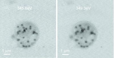
Whole‐mount (WM) platelet preparation followed by transmission electron microscopy (TEM) observation is the standard method currently used to assess dense granule (DG) deficiency (DGD). However, due to the electron‐density‐based contrast mechanism in TEM, other granules such as α‐granules might cause false DG detection. Here, scanning transmission X‐ray microscopy (STXM) was used to identify DGs and minimize false DG detection of human platelets. STXM image stacks of human platelets were collected at the calcium (Ca) L2,3 absorption edge and then converted to optical density maps. Ca distribution maps, obtained by subtracting the optical density maps at the pre‐edge region from those at the post‐edge region, were used to identify DGs based on the Ca richness. DGs were successfully detected using this STXM method without false detection, based on Ca maps for four human platelets. Spectral analysis of granules in human platelets confirmed that DGs contain a richer Ca content than other granules. The Ca distribution maps facilitated more effective DG identification than TEM which might falsely detect DGs. Correct identification of DGs would be important to assess the status of platelets and DG‐related diseases. Therefore, this STXM method is proposed as a promising approach for better DG identification and diagnosis, as a complementary tool to the current WM TEM approach.
中文翻译:

使用扫描透射 X 射线显微镜鉴定人血小板中富含钙的致密颗粒。
全片(WM)血小板制备后进行透射电子显微镜(TEM)观察是目前用于评估致密颗粒(DG)缺陷(DGD)的标准方法。然而,由于 TEM 中基于电子密度的对比机制,其他颗粒(例如 α 颗粒)可能会导致错误的 DG 检测。在这里,扫描透射 X 射线显微镜 (STXM) 用于识别 DG 并最大限度地减少人血小板的错误 DG 检测。在钙 (Ca) L 2,3吸收边缘收集人类血小板的 STXM 图像堆栈,然后转换为光密度图。Ca 分布图是通过从边缘后区域减去边缘前区域的光密度图获得的,用于根据 Ca 丰富度来识别 DG。基于四种人类血小板的 Ca 图谱,使用这种 STXM 方法成功检测到了 DG,没有出现错误检测。对人血小板颗粒的光谱分析证实,DG 比其他颗粒含有更丰富的钙含量。Ca 分布图比 TEM 更有效地识别 DG,而 TEM 可能会错误地检测 DG。正确识别 DG 对于评估血小板状态和 DG 相关疾病非常重要。因此,这种 STXM 方法被认为是一种有前途的方法,可以更好地识别和诊断 DG,作为当前 WM TEM 方法的补充工具。
更新日期:2020-03-16
中文翻译:
使用扫描透射 X 射线显微镜鉴定人血小板中富含钙的致密颗粒。
全片(WM)血小板制备后进行透射电子显微镜(TEM)观察是目前用于评估致密颗粒(DG)缺陷(DGD)的标准方法。然而,由于 TEM 中基于电子密度的对比机制,其他颗粒(例如 α 颗粒)可能会导致错误的 DG 检测。在这里,扫描透射 X 射线显微镜 (STXM) 用于识别 DG 并最大限度地减少人血小板的错误 DG 检测。在钙 (Ca) L 2,3吸收边缘收集人类血小板的 STXM 图像堆栈,然后转换为光密度图。Ca 分布图是通过从边缘后区域减去边缘前区域的光密度图获得的,用于根据 Ca 丰富度来识别 DG。基于四种人类血小板的 Ca 图谱,使用这种 STXM 方法成功检测到了 DG,没有出现错误检测。对人血小板颗粒的光谱分析证实,DG 比其他颗粒含有更丰富的钙含量。Ca 分布图比 TEM 更有效地识别 DG,而 TEM 可能会错误地检测 DG。正确识别 DG 对于评估血小板状态和 DG 相关疾病非常重要。因此,这种 STXM 方法被认为是一种有前途的方法,可以更好地识别和诊断 DG,作为当前 WM TEM 方法的补充工具。


 京公网安备 11010802027423号