当前位置:
X-MOL 学术
›
J. Raman Spectrosc.
›
论文详情
Our official English website, www.x-mol.net, welcomes your
feedback! (Note: you will need to create a separate account there.)
Serial section Raman tomography with 10 times higher depth resolution than confocal Raman microscopy
Journal of Raman Spectroscopy ( IF 2.4 ) Pub Date : 2020-03-23 , DOI: 10.1002/jrs.5878 Thomas Böhm 1, 2 , Riko Moroni 3 , Simon Thiele 1, 2
Journal of Raman Spectroscopy ( IF 2.4 ) Pub Date : 2020-03-23 , DOI: 10.1002/jrs.5878 Thomas Böhm 1, 2 , Riko Moroni 3 , Simon Thiele 1, 2
Affiliation
|
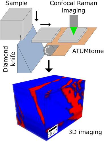
Confocal Raman microscopy enables 3D imaging of various samples solely based on chemical contrast. However, optical artifacts impair resolution and image quality in subsurface imaging. With serial section Raman tomography, we show that serial ultrathin and semithin sectioning by ultramicrotomy can successfully be combined with subsequent confocal Raman imaging. This new 3D Raman imaging technique reaches a depth resolution of up to 100 nm, which is about 10‐fold better than in confocal Raman microscopy. Structurally complex and optically inhomogeneous samples can not only be imaged, but also be used to quantify structural parameters. Serial section Raman tomography is a promising method for materials science and possibly also for life sciences.
中文翻译:

连续切片拉曼断层扫描,其深度分辨率比共聚焦拉曼显微镜高十倍
共焦拉曼显微镜可以仅基于化学对比就可以对各种样品进行3D成像。但是,光学伪影会损害地下成像中的分辨率和图像质量。通过连续切片拉曼断层扫描,我们显示通过超薄切片术进行的连续超薄和半薄切片可以成功地与随后的共聚焦拉曼成像相结合。这项新的3D拉曼成像技术可达到高达100 nm的深度分辨率,这比共聚焦拉曼显微镜要好10倍。结构复杂且光学上不均匀的样本不仅可以成像,还可以用于量化结构参数。串行截面拉曼层析成像是材料科学乃至生命科学的一种有前途的方法。
更新日期:2020-03-23
中文翻译:
连续切片拉曼断层扫描,其深度分辨率比共聚焦拉曼显微镜高十倍
共焦拉曼显微镜可以仅基于化学对比就可以对各种样品进行3D成像。但是,光学伪影会损害地下成像中的分辨率和图像质量。通过连续切片拉曼断层扫描,我们显示通过超薄切片术进行的连续超薄和半薄切片可以成功地与随后的共聚焦拉曼成像相结合。这项新的3D拉曼成像技术可达到高达100 nm的深度分辨率,这比共聚焦拉曼显微镜要好10倍。结构复杂且光学上不均匀的样本不仅可以成像,还可以用于量化结构参数。串行截面拉曼层析成像是材料科学乃至生命科学的一种有前途的方法。


 京公网安备 11010802027423号