当前位置:
X-MOL 学术
›
J. Raman Spectrosc.
›
论文详情
Our official English website, www.x-mol.net, welcomes your
feedback! (Note: you will need to create a separate account there.)
A review of the applications of Raman spectroscopy for breast cancer tissue diagnostic and their histopathological classification of epithelial to mesenchymal transition
Journal of Raman Spectroscopy ( IF 2.4 ) Pub Date : 2019-11-04 , DOI: 10.1002/jrs.5774 Siti Norbaini Sabtu 1 , S.F. Abdul Sani 1 , D.A. Bradley 2, 3 , L.M. Looi 4 , Z. Osman 1
Journal of Raman Spectroscopy ( IF 2.4 ) Pub Date : 2019-11-04 , DOI: 10.1002/jrs.5774 Siti Norbaini Sabtu 1 , S.F. Abdul Sani 1 , D.A. Bradley 2, 3 , L.M. Looi 4 , Z. Osman 1
Affiliation
|
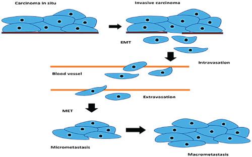
Breast cancer is one of the leading cancers in women worldwide. Notwithstanding the clear advances being made in treatment, early diagnosis of the disease can certainly be expected to reduce morbidity and mortality. With increasing evidence of the role of epithelial to mesenchymal transition (EMT) in tumour progression, early detection of this phenomenon is suggested to be important given that the majority of breast cancer deaths are due to tumour invasion and metastasis. Although histopathology and biomedical imaging techniques continue to be used as standard procedures in breast cancer diagnosis, these techniques have a number of disadvantages, including being time‐consuming, the imaging in particular having attendant limited resolution, sensitivity, and specificity, leading to results that are prone to errors in human interpretation. Due to its rapidity and high specificity, Raman spectroscopy has emerged as a diagnostic tool for breast cancer, useful in identifying malignancy of breast cells, correlated with the EMT phenotype, expressed at the molecular level. Detailed biochemical information from tissue biopsies can also be provided from use of this technique. The use of Raman spectroscopy in breast cancer investigations over the past 10 years and more, including in the study of EMT, is reviewed in the present work also listing the corresponding Raman peaks reported in the literature in seeking to better facilitate identification of peaks of interest.
中文翻译:

拉曼光谱在乳腺癌组织诊断中的应用及其上皮向间质转化的组织病理学分类
乳腺癌是全球女性的主要癌症之一。尽管在治疗方面取得了明显的进步,但是可以肯定地期望疾病的早期诊断可以降低发病率和死亡率。随着越来越多的证据表明上皮到间充质转变(EMT)在肿瘤进展中的作用,鉴于大多数乳腺癌死亡是由于肿瘤的侵袭和转移,因此早期发现这种现象被认为很重要。尽管组织病理学和生物医学成像技术继续被用作乳腺癌诊断的标准程序,但是这些技术具有许多缺点,包括耗时,成像特别是伴随着有限的分辨率,敏感性和特异性,从而导致结果在人类解释中容易出错。由于其快速性和高特异性,拉曼光谱法已成为乳腺癌的诊断工具,可用于在分子水平上鉴定与EMT表型相关的乳腺癌细胞的恶性程度。使用这种技术也可以提供组织活检的详细生化信息。在当前的工作中,对拉曼光谱在过去10年及更长的时间(包括在EMT研究中)在乳腺癌研究中的使用进行了综述,并列出了文献中报道的相应拉曼峰,以寻求更好地促进对感兴趣峰的识别。 。也可以通过使用该技术来提供组织活检的详细生化信息。在当前的工作中,对拉曼光谱在过去10年及更长的时间(包括在EMT研究中)在乳腺癌研究中的使用进行了综述,并列出了文献中报道的相应拉曼峰,以寻求更好地促进对感兴趣峰的识别。 。使用这种技术也可以提供组织活检的详细生化信息。在当前的工作中,对拉曼光谱在过去10年及更长的时间(包括在EMT研究中)在乳腺癌研究中的使用进行了综述,并列出了文献中报道的相应拉曼峰,以寻求更好地促进对感兴趣峰的识别。 。
更新日期:2019-11-04
中文翻译:
拉曼光谱在乳腺癌组织诊断中的应用及其上皮向间质转化的组织病理学分类
乳腺癌是全球女性的主要癌症之一。尽管在治疗方面取得了明显的进步,但是可以肯定地期望疾病的早期诊断可以降低发病率和死亡率。随着越来越多的证据表明上皮到间充质转变(EMT)在肿瘤进展中的作用,鉴于大多数乳腺癌死亡是由于肿瘤的侵袭和转移,因此早期发现这种现象被认为很重要。尽管组织病理学和生物医学成像技术继续被用作乳腺癌诊断的标准程序,但是这些技术具有许多缺点,包括耗时,成像特别是伴随着有限的分辨率,敏感性和特异性,从而导致结果在人类解释中容易出错。由于其快速性和高特异性,拉曼光谱法已成为乳腺癌的诊断工具,可用于在分子水平上鉴定与EMT表型相关的乳腺癌细胞的恶性程度。使用这种技术也可以提供组织活检的详细生化信息。在当前的工作中,对拉曼光谱在过去10年及更长的时间(包括在EMT研究中)在乳腺癌研究中的使用进行了综述,并列出了文献中报道的相应拉曼峰,以寻求更好地促进对感兴趣峰的识别。 。也可以通过使用该技术来提供组织活检的详细生化信息。在当前的工作中,对拉曼光谱在过去10年及更长的时间(包括在EMT研究中)在乳腺癌研究中的使用进行了综述,并列出了文献中报道的相应拉曼峰,以寻求更好地促进对感兴趣峰的识别。 。使用这种技术也可以提供组织活检的详细生化信息。在当前的工作中,对拉曼光谱在过去10年及更长的时间(包括在EMT研究中)在乳腺癌研究中的使用进行了综述,并列出了文献中报道的相应拉曼峰,以寻求更好地促进对感兴趣峰的识别。 。


 京公网安备 11010802027423号