Journal of Neuroradiology ( IF 3.0 ) Pub Date : 2020-03-20 , DOI: 10.1016/j.neurad.2020.03.005 Sylvia M Bardet 1 , Jonathan Cortese 2 , Raphaël Blanc 3 , Charbel Mounayer 4 , Aymeric Rouchaud 4
|
Background
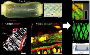
Conventional histological analyses are the gold standard for the study of aneurysms and vascular pathologies in pre-clinical research. Over the past decade, in vivo and ex vivo imaging using multiphoton microscopy have emerged as powerful pre-clinical tools for detailed tissue analyses that can assess morphology, the extracellular matrix (ECM), cell density and vascularisation. Multiphoton microscopy allows for deeper tissue penetration with minor phototoxicity.
Objective
The present study aimed to demonstrate the current status of multimodality imaging, including multiphoton microscopy, for detailed analyses of neo-endothelialisation and ECM evolution after flow-diverter stent (FDS) treatment in an experimental rabbit model of aneurysms.
Methods
Multiphoton microscopy tools for assessing autofluorescence and second harmonic generation (SHG) signals from biological tissues were used to evaluate the endovascular treatment of intracranial aneurysms in an animal model of aneurysms (pig, rabbit). Results from multiphoton microscopy were compared to those from standard histology, electronic and bright field microscopy.
Conclusions
The present study describes novel evaluation modes based on multiphoton microscopy for visualising tissue morphology (e.g., collagen, elastin, and cells) to qualify and quantify the extent of neo-intimal formation of covered arteries and device integration into the arterial wall using a rabbit model of intracranial aneurysms treated with FDS.
中文翻译:
用于治疗动脉瘤的分流支架的临床前评估的多光子显微镜
背景
常规组织学分析是临床前研究中动脉瘤和血管病理学研究的金标准。在过去十年中,使用多光子显微镜进行的体内和体外成像已成为用于详细组织分析的强大的临床前工具,可以评估形态学、细胞外基质 (ECM)、细胞密度和血管形成。多光子显微镜允许以较小的光毒性进行更深的组织渗透。
客观的
本研究旨在证明多模态成像(包括多光子显微镜)的现状,以详细分析实验兔动脉瘤模型中分流支架 (FDS) 治疗后的新内皮化和 ECM 演变。
方法
用于评估来自生物组织的自发荧光和二次谐波生成 (SHG) 信号的多光子显微镜工具用于评估动脉瘤动物模型(猪、兔)中颅内动脉瘤的血管内治疗。将多光子显微镜的结果与标准组织学、电子和明视野显微镜的结果进行比较。
结论
本研究描述了基于多光子显微镜的新型评估模式,用于可视化组织形态(例如,胶原蛋白、弹性蛋白和细胞),以限定和量化覆盖动脉的新内膜形成程度,并使用兔模型将装置整合到动脉壁中用 FDS 治疗的颅内动脉瘤。


 京公网安备 11010802027423号