Photoacoustics ( IF 7.9 ) Pub Date : 2020-03-16 , DOI: 10.1016/j.pacs.2020.100178 Héctor Estrada , Johannes Rebling , Urs Hofmann , Daniel Razansky
|
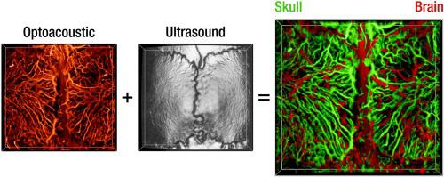
Bone microvasculature plays a paramount role in bone marrow maintenance, development, and hematopoiesis. Studies of calvarian vascular patterns within living mammalian skull with the available intravital microscopy techniques are limited to small scale observations. We developed an optical-resolution optoacoustic microscopy method combined with ultrasound biomicroscopy in order to reveal and discern the intricate networks of calvarian and cerebral vasculature over large fields of view covering majority of the murine calvaria. The vasculature segmentation method is based on an angle-corrected homogeneous model of the rodent skull, generated using simultaneously acquired three-dimensional pulse-echo ultrasound images. The hybrid microscopy design along with the appropriate skull segmentation method enable high throughput studies of a living bone while facilitating correct anatomical interpretation of the vasculature images acquired with optical resolution optoacoustic microscopy.
中文翻译:
结合光声超声显微镜识别颅盖血管微血管网络
骨骼微血管在骨髓维持,发育和造血中起着至关重要的作用。利用活体显微镜技术研究活的哺乳动物颅骨内的颅盖血管模式仅限于小规模观察。我们开发了一种光学分辨率的光声显微镜技术,并将其与超声生物显微镜技术相结合,以揭示和辨别覆盖大部分鼠颅的大视野内的颅骨和脑血管系统的复杂网络。脉管系统分割方法基于啮齿动物头骨的角度校正均匀模型,该模型使用同时获取的三维脉冲回波超声图像生成。


 京公网安备 11010802027423号