当前位置:
X-MOL 学术
›
J. Struct. Biol.
›
论文详情
Our official English website, www.x-mol.net, welcomes your
feedback! (Note: you will need to create a separate account there.)
Pathology of myelinated axons in the PLP-deficient mouse model of spastic paraplegia type 2 revealed by volume imaging using focused ion beam-scanning electron microscopy.
Journal of Structural Biology ( IF 3.0 ) Pub Date : 2020-03-08 , DOI: 10.1016/j.jsb.2020.107492 Anna M Steyer 1 , Torben Ruhwedel 2 , Christos Nardis 1 , Hauke B Werner 3 , Klaus-Armin Nave 3 , Wiebke Möbius 1
Journal of Structural Biology ( IF 3.0 ) Pub Date : 2020-03-08 , DOI: 10.1016/j.jsb.2020.107492 Anna M Steyer 1 , Torben Ruhwedel 2 , Christos Nardis 1 , Hauke B Werner 3 , Klaus-Armin Nave 3 , Wiebke Möbius 1
Affiliation
|
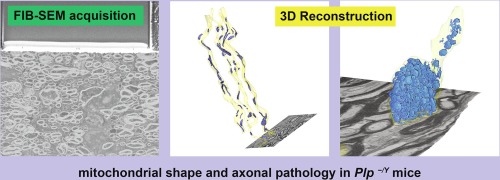
Advances in electron microscopy including improved imaging techniques and state-of-the-art detectors facilitate imaging of larger tissue volumes with electron microscopic resolution. In combination with genetic tools for the generation of mouse mutants this allows assessing the three-dimensional (3D) characteristics of pathological features in disease models. Here we revisited the axonal pathology in the central nervous system of a mouse model of spastic paraplegia type 2, the Plp-/Y mouse. Although PLP is a bona fide myelin protein, the major hallmark of the disease in both SPG2 patients and mouse models are axonal swellings comprising accumulations of numerous organelles including mitochondria, gradually leading to irreversible axonal loss. To assess the number and morphology of axonal mitochondria and the overall myelin preservation we evaluated two sample preparation techniques, chemical fixation or high-pressure freezing and freeze substitution, with respect to the objective of 3D visualization. Both methods allowed visualizing distribution and morphological details of axonal mitochondria. In Plp-/Y mice the number of mitochondria is 2-fold increased along the entire axonal length. Mitochondria are also found in the excessive organelle accumulations within axonal swellings. In addition, organelle accumulations were detected within the myelin sheath and the inner tongue. We find that 3D electron microscopy is required for a comprehensive understanding of the size, content and frequency of axonal swellings, the hallmarks of axonal pathology.
中文翻译:

使用聚焦离子束扫描电子显微镜进行体积成像,揭示了 2 型痉挛性截瘫 PLP 缺陷小鼠模型中有髓轴突的病理学。
电子显微镜的进步,包括改进的成像技术和最先进的探测器,有助于以电子显微镜分辨率对更大的组织体积进行成像。与生成小鼠突变体的遗传工具相结合,可以评估疾病模型中病理特征的三维 (3D) 特征。在这里,我们重新审视了 2 型痉挛性截瘫小鼠模型(Plp-/Y 小鼠)中枢神经系统的轴突病理学。尽管 PLP 是一种真正的髓磷脂蛋白,但 SPG2 患者和小鼠模型中该疾病的主要标志是轴突肿胀,包括包括线粒体在内的众多细胞器的积累,逐渐导致不可逆的轴突损失。为了评估轴突线粒体的数量和形态以及整体髓磷脂保存,我们针对 3D 可视化的目标评估了两种样品制备技术:化学固定或高压冷冻和冷冻替代。两种方法都可以可视化轴突线粒体的分布和形态细节。在 Plp-/Y 小鼠中,整个轴突长度上的线粒体数量增加了 2 倍。线粒体也存在于轴突肿胀内过多的细胞器积累中。此外,在髓鞘和内舌内检测到细胞器积聚。我们发现,需要 3D 电子显微镜来全面了解轴突肿胀的大小、内容和频率,这是轴突病理学的标志。
更新日期:2020-03-26
中文翻译:
使用聚焦离子束扫描电子显微镜进行体积成像,揭示了 2 型痉挛性截瘫 PLP 缺陷小鼠模型中有髓轴突的病理学。
电子显微镜的进步,包括改进的成像技术和最先进的探测器,有助于以电子显微镜分辨率对更大的组织体积进行成像。与生成小鼠突变体的遗传工具相结合,可以评估疾病模型中病理特征的三维 (3D) 特征。在这里,我们重新审视了 2 型痉挛性截瘫小鼠模型(Plp-/Y 小鼠)中枢神经系统的轴突病理学。尽管 PLP 是一种真正的髓磷脂蛋白,但 SPG2 患者和小鼠模型中该疾病的主要标志是轴突肿胀,包括包括线粒体在内的众多细胞器的积累,逐渐导致不可逆的轴突损失。为了评估轴突线粒体的数量和形态以及整体髓磷脂保存,我们针对 3D 可视化的目标评估了两种样品制备技术:化学固定或高压冷冻和冷冻替代。两种方法都可以可视化轴突线粒体的分布和形态细节。在 Plp-/Y 小鼠中,整个轴突长度上的线粒体数量增加了 2 倍。线粒体也存在于轴突肿胀内过多的细胞器积累中。此外,在髓鞘和内舌内检测到细胞器积聚。我们发现,需要 3D 电子显微镜来全面了解轴突肿胀的大小、内容和频率,这是轴突病理学的标志。


 京公网安备 11010802027423号