当前位置:
X-MOL 学术
›
J. Struct. Biol.
›
论文详情
Our official English website, www.x-mol.net, welcomes your
feedback! (Note: you will need to create a separate account there.)
Nanostructure of mouse otoconia.
Journal of Structural Biology ( IF 3.0 ) Pub Date : 2020-03-03 , DOI: 10.1016/j.jsb.2020.107489 Dimitra Athanasiadou 1 , Wenge Jiang 1 , Natalie Reznikov 2 , Alejandro B Rodríguez-Navarro 3 , Roland Kröger 4 , Matthew Bilton 5 , Alicia González-Segura 6 , Yongfeng Hu 7 , Valentin Nelea 1 , Marc D McKee 8
Journal of Structural Biology ( IF 3.0 ) Pub Date : 2020-03-03 , DOI: 10.1016/j.jsb.2020.107489 Dimitra Athanasiadou 1 , Wenge Jiang 1 , Natalie Reznikov 2 , Alejandro B Rodríguez-Navarro 3 , Roland Kröger 4 , Matthew Bilton 5 , Alicia González-Segura 6 , Yongfeng Hu 7 , Valentin Nelea 1 , Marc D McKee 8
Affiliation
|
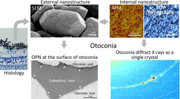
Mammalian otoconia of the inner ear vestibular apparatus are calcium carbonate-containing mineralized structures critical for maintaining balance and detecting linear acceleration. The mineral phase of otoconia is calcite, which coherently diffracts X-rays much like a single-crystal. Otoconia contain osteopontin (OPN), a mineral-binding protein influencing mineralization processes in bones, teeth and avian eggshells, for example, and in pathologic mineral deposits. Here we describe mineral nanostructure and the distribution of OPN in mouse otoconia. Scanning electron microscopy and atomic force microscopy of intact and cleaved mouse otoconia revealed an internal nanostructure (~50 nm). Transmission electron microscopy and electron tomography of focused ion beam-prepared sections of otoconia confirmed this mineral nanostructure, and identified even smaller (~10 nm) nanograin dimensions. X-ray diffraction of mature otoconia (8-day-old mice) showed crystallite size in a similar range (73 nm and smaller). Raman and X-ray absorption spectroscopy - both methods being sensitive to the detection of crystalline and amorphous forms in the sample - showed no evidence of amorphous calcium carbonate in these mature otoconia. Scanning and transmission electron microscopy combined with colloidal-gold immunolabeling for OPN revealed that this protein was located at the surface of the otoconia, correlating with a site where surface nanostructure was observed. OPN addition to calcite growing in vitro produced similar surface nanostructure. These findings provide details on the composition and nanostructure of mammalian otoconia, and suggest that while OPN may influence surface rounding and surface nanostructure in otoconia, other incorporated proteins (also possibly including OPN) likely participate in creating internal nanostructure.
中文翻译:

小鼠耳垢的纳米结构。
内耳前庭装置的哺乳动物耳垢是含碳酸钙的矿化结构,对于保持平衡和检测线性加速度至关重要。耳石症的矿物相是方解石,它像单晶一样相干地衍射X射线。Otoconia含有骨桥蛋白(OPN),这是一种矿物质结合蛋白,可影响例如骨骼,牙齿和禽蛋壳以及病理性矿物质沉积物中的矿化过程。在这里,我们描述了矿物质的纳米结构和OPN在小鼠耳菌病中的分布。完整和裂解的小鼠耳垢的扫描电子显微镜和原子力显微镜显示内部纳米结构(〜50 nm)。透射电镜和电子断层扫描仪对聚焦的离子束制备的耳垢进行了确认,证实了这种矿物纳米结构,并确定了更小的(〜10 nm)纳米颗粒尺寸。成熟的耳菌病(8天大的小鼠)的X射线衍射显示出微晶尺寸在相似的范围内(73纳米或更小)。拉曼和X射线吸收光谱法-两种方法都对检测样品中的结晶形式和无定形形式均很敏感-在这些成熟的耳菌病中没有发现无定形碳酸钙的证据。扫描和透射电子显微镜结合胶体金免疫标记的OPN表明,该蛋白位于otoconia的表面,与观察到表面纳米结构的位置相关。OPN除了在方解石中生长外,产生了相似的表面纳米结构。这些发现提供了有关哺乳动物耳垢的组成和纳米结构的详细信息,
更新日期:2020-03-26
中文翻译:
小鼠耳垢的纳米结构。
内耳前庭装置的哺乳动物耳垢是含碳酸钙的矿化结构,对于保持平衡和检测线性加速度至关重要。耳石症的矿物相是方解石,它像单晶一样相干地衍射X射线。Otoconia含有骨桥蛋白(OPN),这是一种矿物质结合蛋白,可影响例如骨骼,牙齿和禽蛋壳以及病理性矿物质沉积物中的矿化过程。在这里,我们描述了矿物质的纳米结构和OPN在小鼠耳菌病中的分布。完整和裂解的小鼠耳垢的扫描电子显微镜和原子力显微镜显示内部纳米结构(〜50 nm)。透射电镜和电子断层扫描仪对聚焦的离子束制备的耳垢进行了确认,证实了这种矿物纳米结构,并确定了更小的(〜10 nm)纳米颗粒尺寸。成熟的耳菌病(8天大的小鼠)的X射线衍射显示出微晶尺寸在相似的范围内(73纳米或更小)。拉曼和X射线吸收光谱法-两种方法都对检测样品中的结晶形式和无定形形式均很敏感-在这些成熟的耳菌病中没有发现无定形碳酸钙的证据。扫描和透射电子显微镜结合胶体金免疫标记的OPN表明,该蛋白位于otoconia的表面,与观察到表面纳米结构的位置相关。OPN除了在方解石中生长外,产生了相似的表面纳米结构。这些发现提供了有关哺乳动物耳垢的组成和纳米结构的详细信息,


 京公网安备 11010802027423号