当前位置:
X-MOL 学术
›
Adv. Funct. Mater.
›
论文详情
Our official English website, www.x-mol.net, welcomes your
feedback! (Note: you will need to create a separate account there.)
Understanding the In Vivo Fate of Advanced Materials by Imaging
Advanced Functional Materials ( IF 18.5 ) Pub Date : 2020-04-06 , DOI: 10.1002/adfm.201910369 Ran Li 1 , Thomas S C Ng 1 , Michelle A Garlin 1 , Ralph Weissleder 1, 2, 3 , Miles A Miller 1, 2
Advanced Functional Materials ( IF 18.5 ) Pub Date : 2020-04-06 , DOI: 10.1002/adfm.201910369 Ran Li 1 , Thomas S C Ng 1 , Michelle A Garlin 1 , Ralph Weissleder 1, 2, 3 , Miles A Miller 1, 2
Affiliation
|
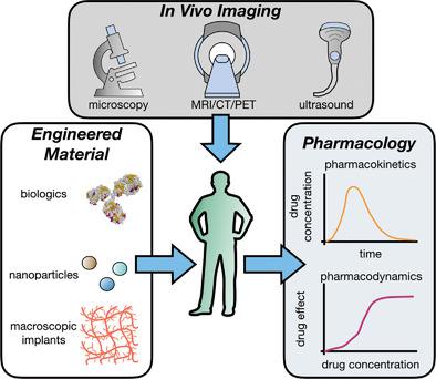
Engineered materials are ubiquitous in biomedical applications ranging from systemic drug delivery systems to orthopedic implants, and their actions unfold across multiple time‐ and length‐scales. The efficacy and safety of biologics, nanomaterials, and macroscopic implants are all dictated by the same general principles of pharmacology as applied to small molecule drugs, comprising how the body affects materials (pharmacokinetics, PK) and conversely how materials affect the body (pharmacodynamics, PD). Imaging technologies play an increasingly insightful role in monitoring both of these processes, often simultaneously: translational macroscopic imaging modalities such as magnetic resonance imaging and positron emission tomography/computed tomography offer whole‐body quantitation of biodistribution and structural or molecular response, while ex vivo approaches and optical imaging via in vivo (intravital) microscopy reveal behaviors at subcellular resolution. In this review, the authors survey developments in imaging the in situ behavior of systemically and locally administered materials, with a particular focus on using microscopy to understand transport, target engagement, and downstream host responses at a single‐cell level. The themes of microenvironmental influence, controlled drug release, on‐target molecular action, and immune response, especially as mediated by macrophages and other myeloid cells, are examined. Finally, the future directions of how new imaging technologies may propel efficient clinical translation of next‐generation therapeutics and medical devices are proposed.
中文翻译:

通过成像了解先进材料的体内命运
工程材料在生物医学应用中无处不在,从全身药物输送系统到骨科植入物,它们的作用在多个时间和长度尺度上展开。生物制品、纳米材料和宏观植入物的功效和安全性都由应用于小分子药物的相同药理学一般原理决定,包括身体如何影响材料(药代动力学,PK)以及相反材料如何影响身体(药效学, PD)。成像技术在监测这两个过程中发挥着越来越深刻的作用,通常是同时进行:平移宏观成像模式,如磁共振成像和正电子发射断层扫描/计算机断层扫描,提供生物分布和结构或分子反应的全身定量,而离体方法通过体内(活体)显微镜的光学成像揭示了亚细胞分辨率的行为。在这篇综述中,作者调查了全身和局部施用材料的原位行为成像的进展,特别关注使用显微镜来了解单细胞水平的运输、目标参与和下游宿主反应。研究的主题包括微环境影响、受控药物释放、靶向分子作用和免疫反应,特别是由巨噬细胞和其他骨髓细胞介导的免疫反应。最后,提出了新成像技术如何推动下一代治疗方法和医疗设备高效临床转化的未来方向。
更新日期:2020-04-06
中文翻译:
通过成像了解先进材料的体内命运
工程材料在生物医学应用中无处不在,从全身药物输送系统到骨科植入物,它们的作用在多个时间和长度尺度上展开。生物制品、纳米材料和宏观植入物的功效和安全性都由应用于小分子药物的相同药理学一般原理决定,包括身体如何影响材料(药代动力学,PK)以及相反材料如何影响身体(药效学, PD)。成像技术在监测这两个过程中发挥着越来越深刻的作用,通常是同时进行:平移宏观成像模式,如磁共振成像和正电子发射断层扫描/计算机断层扫描,提供生物分布和结构或分子反应的全身定量,而离体方法通过体内(活体)显微镜的光学成像揭示了亚细胞分辨率的行为。在这篇综述中,作者调查了全身和局部施用材料的原位行为成像的进展,特别关注使用显微镜来了解单细胞水平的运输、目标参与和下游宿主反应。研究的主题包括微环境影响、受控药物释放、靶向分子作用和免疫反应,特别是由巨噬细胞和其他骨髓细胞介导的免疫反应。最后,提出了新成像技术如何推动下一代治疗方法和医疗设备高效临床转化的未来方向。


 京公网安备 11010802027423号