当前位置:
X-MOL 学术
›
Sci. Total Environ.
›
论文详情
Our official English website, www.x-mol.net, welcomes your
feedback! (Note: you will need to create a separate account there.)
Methyl mercury (MeHg) in vitro exposure alters mitogen-induced lymphocyte proliferation and cytokine expression in Steller sea lion (Eumetopias jubatus) pups.
Science of the Total Environment ( IF 8.2 ) Pub Date : 2020-04-06 , DOI: 10.1016/j.scitotenv.2020.138308 Milton Levin 1 , Lindsay Jasperse 1 , Jean-Pierre Desforges 2 , Todd O'Hara 3 , Lorrie Rea 4 , J Margaret Castellini 5 , John M Maniscalco 6 , Brian Fadely 7 , Mandy Keogh 8
Science of the Total Environment ( IF 8.2 ) Pub Date : 2020-04-06 , DOI: 10.1016/j.scitotenv.2020.138308 Milton Levin 1 , Lindsay Jasperse 1 , Jean-Pierre Desforges 2 , Todd O'Hara 3 , Lorrie Rea 4 , J Margaret Castellini 5 , John M Maniscalco 6 , Brian Fadely 7 , Mandy Keogh 8
Affiliation
|
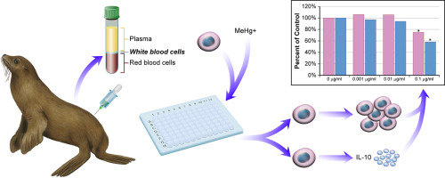
Steller sea lions (Eumetopias jubatus, SSLs) are managed as two distinct population segments within U.S. waters: the endangered western distinct population segment and the recently delisted eastern distinct population segment. Recent studies reported concentrations of mercury in several tissues collected from young SSLs in the Aleutian Islands that were at or above concentrations found to negatively impact health in other fish-eating mammals. However, there are limited studies which have investigated the range of mercury concentrations that may negatively influence the SSL immune system. This study assessed relationships between methyl mercury (MeHg+) concentrations and two immune functions, lymphocyte proliferation and cytokine expression. Peripheral blood mononuclear cells (PBMCs) were isolated and cryopreserved from pups on three rookeries within the western distinct population segment: Chiswell Island, Ulak, and Agattu Islands. Lymphocyte proliferation and cytokine expression were assessed in vitro using thawed PBMCs with exposure to MeHg+ (unexposed control, 0.001, 0.01, and 0.1 μg/ml). Lymphocyte proliferation was measured without and with stimulation with a T cell mitogen (ConA) and B cell mitogen (LPS) and the concentration of cytokines was measured in the cell culture supernatant (with and without ConA or LPS). Spontaneous lymphocyte proliferation was significantly increased at 0.01 and 0.1 μg/ml. T lymphocyte proliferation was significantly increased at 0.001 μg/ml and 0.1 μg/ml, while B lymphocyte proliferation was decreased at 0.1 μg/ml. Cytokine concentrations for INFγ, IL-10, IL-6, and TNFα were reduced at 0.1 μg/ml upon either T or B cell mitogen stimulation, with the exception for IL-10, where 0.1 μg/ml reduced IL-10 concentration compared to unstimulated cells. These data suggest immune functions were affected by MeHg+ exposure requiring in vivo follow up investigations. The observed modulation of immune functions is of concern as any toxicant-induced modulation may adversely affect the health of individuals, particularly younger animals undergoing periods of critical development.
中文翻译:

甲基汞(MeHg)的体外暴露改变了Steller海狮(Eumetopias jubatus)幼仔中有丝分裂原诱导的淋巴细胞增殖和细胞因子表达。
斯特勒海狮(Eumetopias jubatus,SSLs)作为美国水域中两个不同的种群分类进行管理:濒临灭绝的西部不同种群分类和最近被除名的东部不同种群分类。最近的研究报告说,从阿留申群岛的年轻SSL收集的一些组织中的汞浓度等于或高于对其他食鱼哺乳动物的健康产生不利影响的浓度。但是,很少有研究调查可能对SSL免疫系统产生负面影响的汞浓度范围。这项研究评估了甲基汞(MeHg +)浓度与两种免疫功能,淋巴细胞增殖和细胞因子表达之间的关系。分离出外周血单核细胞(PBMC),并从西部独特人群(Chiswell Island,Ulak和Agattu Islands)的三个群中的幼犬中冷冻保存。使用解冻的PBMC暴露于MeHg +(未暴露的对照,0.001、0.01和0.1μg/ ml)在体外评估淋巴细胞的增殖和细胞因子的表达。在没有和有T细胞促细胞分裂剂(ConA)和B细胞促细胞分裂剂(LPS)刺激的情况下测量淋巴细胞增殖,并在细胞培养上清液中(有和没有ConA或LPS)测量细胞因子的浓度。自发性淋巴细胞增殖以0.01和0.1μg/ ml显着增加。T淋巴细胞增殖分别以0.001μg/ ml和0.1μg/ ml显着增加,而B淋巴细胞增殖以0.1μg/ ml降低。INFγ的细胞因子浓度,T或B细胞有丝分裂原刺激后,IL-10,IL-6和TNFα降低了0.1μg/ ml,IL-10除外,与未刺激的细胞相比,IL-10降低了0.1μg/ ml。这些数据表明免疫功能受MeHg +暴露影响,需要进行体内随访研究。观察到的免疫功能调节是令人关注的,因为任何由毒物引起的调节都可能对个体的健康产生不利影响,特别是处于关键发育时期的年幼动物。
更新日期:2020-04-06
中文翻译:
甲基汞(MeHg)的体外暴露改变了Steller海狮(Eumetopias jubatus)幼仔中有丝分裂原诱导的淋巴细胞增殖和细胞因子表达。
斯特勒海狮(Eumetopias jubatus,SSLs)作为美国水域中两个不同的种群分类进行管理:濒临灭绝的西部不同种群分类和最近被除名的东部不同种群分类。最近的研究报告说,从阿留申群岛的年轻SSL收集的一些组织中的汞浓度等于或高于对其他食鱼哺乳动物的健康产生不利影响的浓度。但是,很少有研究调查可能对SSL免疫系统产生负面影响的汞浓度范围。这项研究评估了甲基汞(MeHg +)浓度与两种免疫功能,淋巴细胞增殖和细胞因子表达之间的关系。分离出外周血单核细胞(PBMC),并从西部独特人群(Chiswell Island,Ulak和Agattu Islands)的三个群中的幼犬中冷冻保存。使用解冻的PBMC暴露于MeHg +(未暴露的对照,0.001、0.01和0.1μg/ ml)在体外评估淋巴细胞的增殖和细胞因子的表达。在没有和有T细胞促细胞分裂剂(ConA)和B细胞促细胞分裂剂(LPS)刺激的情况下测量淋巴细胞增殖,并在细胞培养上清液中(有和没有ConA或LPS)测量细胞因子的浓度。自发性淋巴细胞增殖以0.01和0.1μg/ ml显着增加。T淋巴细胞增殖分别以0.001μg/ ml和0.1μg/ ml显着增加,而B淋巴细胞增殖以0.1μg/ ml降低。INFγ的细胞因子浓度,T或B细胞有丝分裂原刺激后,IL-10,IL-6和TNFα降低了0.1μg/ ml,IL-10除外,与未刺激的细胞相比,IL-10降低了0.1μg/ ml。这些数据表明免疫功能受MeHg +暴露影响,需要进行体内随访研究。观察到的免疫功能调节是令人关注的,因为任何由毒物引起的调节都可能对个体的健康产生不利影响,特别是处于关键发育时期的年幼动物。


 京公网安备 11010802027423号