当前位置:
X-MOL 学术
›
Acta Biomater.
›
论文详情
Our official English website, www.x-mol.net, welcomes your
feedback! (Note: you will need to create a separate account there.)
Cell encapsulation spatially alters crosslink density of poly(ethylene glycol) hydrogels formed from free-radical polymerizations.
Acta Biomaterialia ( IF 9.4 ) Pub Date : 2020-04-05 , DOI: 10.1016/j.actbio.2020.03.033 Stanley Chu 1 , Mollie M Maples 1 , Stephanie J Bryant 2
Acta Biomaterialia ( IF 9.4 ) Pub Date : 2020-04-05 , DOI: 10.1016/j.actbio.2020.03.033 Stanley Chu 1 , Mollie M Maples 1 , Stephanie J Bryant 2
Affiliation
|
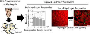
Photopolymerizable poly(ethylene glycol) (PEG) hydrogels are a promising platform for chondrocyte encapsulation and cartilage tissue engineering. This study demonstrates that during the process of encapsulation, chondrocytes alter the formation of PEG hydrogels leading to a reduction in the bulk and local hydrogel crosslink density. Freshly isolated chondrocytes were shown to interact with hydrogel precursors, in part through thiol-mediated events between dithiol crosslinkers and cell surface free thiols, depleting crosslinker concentration and causing a reduction in the bulk hydrogel crosslink density. This effect was more pronounced with increasing cell density at the time of encapsulation. Encapsulation of chondrocytes in fluorescently labeled hydrogels exhibited a gradient in hydrogel density around the cell, which was abrogated by treatment of the cells with the antioxidant estradiol prior to encapsulation. This gradient led to spatial variations in the degradation behavior of a hydrolytically degradable PEG hydrogel, creating regions devoid of hydrogel surrounding cells. Collectively, findings from this study indicate that the antioxidant defense mechanisms in chondrocytes alter the resultant properties of PEG hydrogels formed by free-radical polymerizations. These interactions will have a significant impact on tissue engineering, affecting the local microenvironment around cells and how tissue grows within the hydrogels. STATEMENT OF SIGNIFICANCE: Cell encapsulations in synthetic hydrogels formed by free-radical polymerizations offer numerous benefits for tissue engineering. Herein, we studied cartilage cells and identified that during encapsulation, cells interfered with hydrogel formation through two distinct mechanisms. Thiol-mediated events between monomers led to monomer depletion and a lower crosslinked hydrogel. Cells' antioxidant defense mechanisms interfered with free-radicals and inhibited hydrogel formation near the cell. These cell-mediated effects led to softer hydrogels and created unique hydrogel degradations patterns causing rapid degradation around the cells. The latter has benefits for tissue engineering, where these regions provide space for tissue growth. Overall, this study demonstrates that cells play a key role in how the hydrogel structure forms when cells are present.
中文翻译:

细胞封装在空间上改变了自由基聚合形成的聚乙二醇水凝胶的交联密度。
光聚合聚乙二醇(PEG)水凝胶是软骨细胞封装和软骨组织工程的一个有前途的平台。这项研究表明,在封装过程中,软骨细胞改变了 PEG 水凝胶的形成,导致体积和局部水凝胶交联密度降低。新分离的软骨细胞被证明与水凝胶前体相互作用,部分是通过二硫醇交联剂和细胞表面游离硫醇之间的硫醇介导的事件,耗尽交联剂浓度并导致整体水凝胶交联密度降低。随着封装时细胞密度的增加,这种效应更加明显。将软骨细胞封装在荧光标记的水凝胶中,在细胞周围表现出水凝胶密度梯度,在封装之前用抗氧化剂雌二醇处理细胞可以消除这种梯度。这种梯度导致可水解降解的 PEG 水凝胶的降解行为发生空间变化,从而形成细胞周围没有水凝胶的区域。总的来说,这项研究的结果表明,软骨细胞中的抗氧化防御机制改变了自由基聚合形成的 PEG 水凝胶的最终特性。这些相互作用将对组织工程产生重大影响,影响细胞周围的局部微环境以及组织在水凝胶内的生长方式。意义声明:通过自由基聚合形成的合成水凝胶中的细胞封装为组织工程提供了许多好处。在此,我们研究了软骨细胞,并发现在封装过程中,细胞通过两种不同的机制干扰水凝胶的形成。 单体之间的硫醇介导的事件导致单体耗尽和交联度较低的水凝胶。细胞的抗氧化防御机制干扰自由基并抑制细胞附近水凝胶的形成。这些细胞介导的效应导致水凝胶变软,并产生独特的水凝胶降解模式,导致细胞周围快速降解。后者对组织工程有好处,这些区域为组织生长提供了空间。总的来说,这项研究表明,当细胞存在时,细胞在水凝胶结构的形成中发挥着关键作用。
更新日期:2020-04-05
中文翻译:
细胞封装在空间上改变了自由基聚合形成的聚乙二醇水凝胶的交联密度。
光聚合聚乙二醇(PEG)水凝胶是软骨细胞封装和软骨组织工程的一个有前途的平台。这项研究表明,在封装过程中,软骨细胞改变了 PEG 水凝胶的形成,导致体积和局部水凝胶交联密度降低。新分离的软骨细胞被证明与水凝胶前体相互作用,部分是通过二硫醇交联剂和细胞表面游离硫醇之间的硫醇介导的事件,耗尽交联剂浓度并导致整体水凝胶交联密度降低。随着封装时细胞密度的增加,这种效应更加明显。将软骨细胞封装在荧光标记的水凝胶中,在细胞周围表现出水凝胶密度梯度,在封装之前用抗氧化剂雌二醇处理细胞可以消除这种梯度。这种梯度导致可水解降解的 PEG 水凝胶的降解行为发生空间变化,从而形成细胞周围没有水凝胶的区域。总的来说,这项研究的结果表明,软骨细胞中的抗氧化防御机制改变了自由基聚合形成的 PEG 水凝胶的最终特性。这些相互作用将对组织工程产生重大影响,影响细胞周围的局部微环境以及组织在水凝胶内的生长方式。意义声明:通过自由基聚合形成的合成水凝胶中的细胞封装为组织工程提供了许多好处。在此,我们研究了软骨细胞,并发现在封装过程中,细胞通过两种不同的机制干扰水凝胶的形成。 单体之间的硫醇介导的事件导致单体耗尽和交联度较低的水凝胶。细胞的抗氧化防御机制干扰自由基并抑制细胞附近水凝胶的形成。这些细胞介导的效应导致水凝胶变软,并产生独特的水凝胶降解模式,导致细胞周围快速降解。后者对组织工程有好处,这些区域为组织生长提供了空间。总的来说,这项研究表明,当细胞存在时,细胞在水凝胶结构的形成中发挥着关键作用。


 京公网安备 11010802027423号