当前位置:
X-MOL 学术
›
Biotechnol. Bioeng.
›
论文详情
Our official English website, www.x-mol.net, welcomes your
feedback! (Note: you will need to create a separate account there.)
Quantification of the HIV-1 virus-like particle production process by super-resolution imaging: From VLP budding to nanoparticle analysis.
Biotechnology and Bioengineering ( IF 3.5 ) Pub Date : 2020-04-03 , DOI: 10.1002/bit.27345 Irene González-Domínguez 1 , Eduard Puente-Massaguer 1 , Laura Cervera 1 , Francesc Gòdia 1
Biotechnology and Bioengineering ( IF 3.5 ) Pub Date : 2020-04-03 , DOI: 10.1002/bit.27345 Irene González-Domínguez 1 , Eduard Puente-Massaguer 1 , Laura Cervera 1 , Francesc Gòdia 1
Affiliation

|
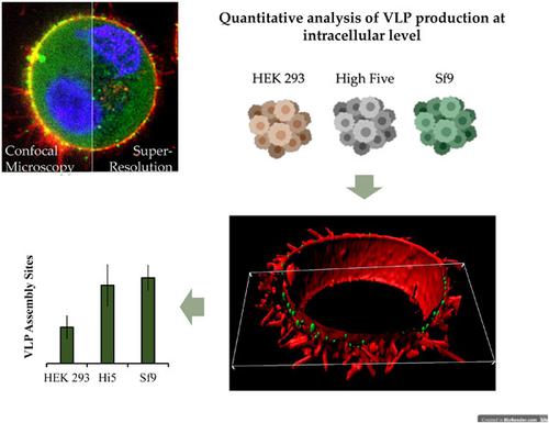
Virus‐like particles (VLPs) offer great promise in the field of nanomedicine. Enveloped VLPs are a class of these nanoparticles and their production process occurs by a budding process, which is known to be the most critical step at intracellular level. In this study, we developed a novel imaging method based on super‐resolution fluorescence microscopy (SRFM) to assess the generation of VLPs in living cells. This methodology was applied to study the production of Gag VLPs in three animal cell platforms of reference: HEK 293‐transient gene expression (TGE), High Five‐baculovirus expression vector system (BEVS) and Sf9‐BEVS. Quantification of the number of VLP assembly sites per cell ranged from 500 to 3,000 in the different systems evaluated. Although the BEVS was superior in terms of Gag polyprotein expression, the HEK 293‐TGE platform was more efficient regarding the assembly of Gag as VLPs. This was translated into higher levels of non‐assembled Gag monomer in BEVS harvested supernatants. Furthermore, the presence of contaminating nanoparticles was evidenced in all three systems, specifically in High Five cells. The SRFM‐based method here developed was also successfully applied to measure the concentration of VLPs in crude supernatants. The lipid membrane of VLPs and the presence of nucleic acids alongside these nanoparticles could also be detected using common staining procedures. Overall, a complete picture of the VLP production process was achieved in these three production platforms. The robustness and sensitivity of this new approach broaden the applicability of SRFM toward the development of new detection, diagnosis and quantification methods based on confocal microscopy in living systems.
中文翻译:

通过超分辨率成像量化 HIV-1 病毒样颗粒生产过程:从 VLP 出芽到纳米颗粒分析。
病毒样颗粒(VLP)在纳米医学领域提供了广阔的前景。包膜 VLP 是这类纳米颗粒中的一类,它们的生产过程是通过出芽过程发生的,这是细胞内水平最关键的步骤。在这项研究中,我们开发了一种基于超分辨率荧光显微镜 (SRFM) 的新型成像方法来评估活细胞中 VLP 的产生。该方法用于研究在三种动物细胞参考平台中 Gag VLP 的产生:HEK 293 瞬时基因表达 (TGE)、高五杆状病毒表达载体系统 (BEVS) 和 Sf9-BEVS。在评估的不同系统中,每个细胞的 VLP 组装位点数量的量化范围为 500 到 3,000。尽管 BEVS 在 Gag 多蛋白表达方面优于 HEK 293-TGE 平台在将 Gag 组装为 VLP 方面效率更高。这被转化为 BEVS 收获的上清液中更高水平的非组装 Gag 单体。此外,在所有三个系统中都证明了污染纳米颗粒的存在,特别是在 High Five 细胞中。这里开发的基于 SRFM 的方法也成功应用于测量粗上清液中 VLP 的浓度。VLP 的脂质膜和这些纳米颗粒旁边的核酸也可以使用常见的染色程序进行检测。总体而言,在这三个生产平台上实现了 VLP 生产过程的完整图景。这种新方法的稳健性和灵敏度拓宽了 SRFM 对新检测开发的适用性,
更新日期:2020-04-03
中文翻译:

通过超分辨率成像量化 HIV-1 病毒样颗粒生产过程:从 VLP 出芽到纳米颗粒分析。
病毒样颗粒(VLP)在纳米医学领域提供了广阔的前景。包膜 VLP 是这类纳米颗粒中的一类,它们的生产过程是通过出芽过程发生的,这是细胞内水平最关键的步骤。在这项研究中,我们开发了一种基于超分辨率荧光显微镜 (SRFM) 的新型成像方法来评估活细胞中 VLP 的产生。该方法用于研究在三种动物细胞参考平台中 Gag VLP 的产生:HEK 293 瞬时基因表达 (TGE)、高五杆状病毒表达载体系统 (BEVS) 和 Sf9-BEVS。在评估的不同系统中,每个细胞的 VLP 组装位点数量的量化范围为 500 到 3,000。尽管 BEVS 在 Gag 多蛋白表达方面优于 HEK 293-TGE 平台在将 Gag 组装为 VLP 方面效率更高。这被转化为 BEVS 收获的上清液中更高水平的非组装 Gag 单体。此外,在所有三个系统中都证明了污染纳米颗粒的存在,特别是在 High Five 细胞中。这里开发的基于 SRFM 的方法也成功应用于测量粗上清液中 VLP 的浓度。VLP 的脂质膜和这些纳米颗粒旁边的核酸也可以使用常见的染色程序进行检测。总体而言,在这三个生产平台上实现了 VLP 生产过程的完整图景。这种新方法的稳健性和灵敏度拓宽了 SRFM 对新检测开发的适用性,


 京公网安备 11010802027423号