当前位置:
X-MOL 学术
›
Colloids Surf. B Biointerfaces
›
论文详情
Our official English website, www.x-mol.net, welcomes your feedback! (Note: you will need to create a separate account there.)
Electrospun poly(vinyl alcohol)/reduced graphene oxide nanofibrous scaffolds for skin tissue engineering.
Colloids and Surfaces B: Biointerfaces ( IF 5.8 ) Pub Date : 2020-04-02 , DOI: 10.1016/j.colsurfb.2020.110994 Kannan Badri Narayanan 1 , Gyu Tae Park 1 , Sung Soo Han 1
Colloids and Surfaces B: Biointerfaces ( IF 5.8 ) Pub Date : 2020-04-02 , DOI: 10.1016/j.colsurfb.2020.110994 Kannan Badri Narayanan 1 , Gyu Tae Park 1 , Sung Soo Han 1
Affiliation
|
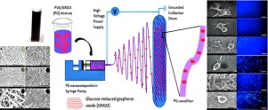
Graphene is composed of a two-dimensional (2D) layer of carbon atoms arranged in a honeycomb lattice configuration. In this paper, we adopted a green synthetic method of producing reduced graphene oxide using glucose as a reducing and stabilizing agent. We also investigated the fabrication of electrospun nanofibers of glucose-reduced graphene oxide (GRGO) (0-1.0 wt%) reinforced with polyvinyl alcohol (PVA) as (PG) scaffolds, and chemically crosslinked with acidic glutaraldehyde (GA) in acetone medium to mimic the extracellular matrix (ECM) for skin tissue engineering applications. These PG scaffolds were evaluated for morphology, mechanical strength, surface wettability, thermal properties, hemocompatibility, and biocompatibility. Field emission-scanning electron microscopy (FE-SEM) revealed an increase in the thickness of nanofibers in PG scaffolds with an increase in the concentration of GRGO. X-ray diffraction and attenuated total reflectance-infrared and Raman spectra showed the GRGO was incorporated in the PVA nanofibrous matrix. As the concentration of GRGO was increased in PG scaffolds, tensile strengths and elongations at break decreased, whereas thermal properties increased. The biological activities of PG scaffolds were evaluated using in vitro hemolysis, using CCD-986Sk (a human skin fibroblast cell line) viability and proliferation assays, and by live/dead cell imaging. Results showed GRGO inclusion in PVA nanofibers caused a slight hydrophilic to hydrophobic shift. PG scaffolds did not cause hemolysis of red blood cells even at a GRGO loading of 1.0 wt%, and PG-1.0 scaffold (with a GRGO loading of 1.0 wt%) exhibited excellent compatibility with fibroblasts and significantly increased metabolic activity after culture for 21 days as compared with PG-0 controls. DAPI staining and live/dead imaging assays showed that all PG scaffolds increased fibroblast proliferation and viability, indicating the potential for skin tissue engineering applications.
中文翻译:

用于皮肤组织工程的静电纺丝聚乙烯醇/还原氧化石墨烯纳米纤维支架。
石墨烯由以蜂窝晶格配置排列的二维(2D)碳原子层组成。在本文中,我们采用了一种绿色合成方法,以葡萄糖为还原稳定剂来生产氧化石墨烯。我们还研究了用聚乙烯醇(PVA)作为(PG)支架增强的葡萄糖还原的氧化石墨烯(GRGO)(0-1.0 wt%)的电纺纳米纤维的制备,并在丙酮介质中与酸性戊二醛(GA)化学交联成模拟细胞外基质(ECM),用于皮肤组织工程应用。评价这些PG支架的形态,机械强度,表面润湿性,热性质,血液相容性和生物相容性。场发射扫描电子显微镜(FE-SEM)显示,PG支架中纳米纤维的厚度随GRGO浓度的增加而增加。X射线衍射和衰减的全反射红外光谱和拉曼光谱表明,GRGO被掺入到PVA纳米纤维基质中。随着PG支架中GRGO浓度的增加,抗张强度和断裂伸长率降低,而热性能提高。使用体外溶血,使用CCD-986Sk(人皮肤成纤维细胞系)生存力和增殖测定以及活/死细胞成像来评估PG支架的生物学活性。结果表明GRGO包含在PVA纳米纤维中会引起轻微的亲水性至疏水性转变。即使在GRGO含量为1.0 wt%的情况下,PG支架也不会引起红细胞的溶血,PG-1.0和PG-1.0支架(GRGO含量为1.0重量%)与成纤维细胞表现出优异的相容性,与PG-0对照相比,培养21天后其代谢活性显着增加。DAPI染色和活/死成像分析表明,所有PG支架均可增加成纤维细胞的增殖和生存能力,表明皮肤组织工程应用的潜力。
更新日期:2020-04-03
中文翻译:
用于皮肤组织工程的静电纺丝聚乙烯醇/还原氧化石墨烯纳米纤维支架。
石墨烯由以蜂窝晶格配置排列的二维(2D)碳原子层组成。在本文中,我们采用了一种绿色合成方法,以葡萄糖为还原稳定剂来生产氧化石墨烯。我们还研究了用聚乙烯醇(PVA)作为(PG)支架增强的葡萄糖还原的氧化石墨烯(GRGO)(0-1.0 wt%)的电纺纳米纤维的制备,并在丙酮介质中与酸性戊二醛(GA)化学交联成模拟细胞外基质(ECM),用于皮肤组织工程应用。评价这些PG支架的形态,机械强度,表面润湿性,热性质,血液相容性和生物相容性。场发射扫描电子显微镜(FE-SEM)显示,PG支架中纳米纤维的厚度随GRGO浓度的增加而增加。X射线衍射和衰减的全反射红外光谱和拉曼光谱表明,GRGO被掺入到PVA纳米纤维基质中。随着PG支架中GRGO浓度的增加,抗张强度和断裂伸长率降低,而热性能提高。使用体外溶血,使用CCD-986Sk(人皮肤成纤维细胞系)生存力和增殖测定以及活/死细胞成像来评估PG支架的生物学活性。结果表明GRGO包含在PVA纳米纤维中会引起轻微的亲水性至疏水性转变。即使在GRGO含量为1.0 wt%的情况下,PG支架也不会引起红细胞的溶血,PG-1.0和PG-1.0支架(GRGO含量为1.0重量%)与成纤维细胞表现出优异的相容性,与PG-0对照相比,培养21天后其代谢活性显着增加。DAPI染色和活/死成像分析表明,所有PG支架均可增加成纤维细胞的增殖和生存能力,表明皮肤组织工程应用的潜力。


 京公网安备 11010802027423号